


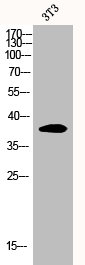
Figure 1. Western blot analysis of EIF2S1 using anti-EIF2S1 antibody (P04387). Electrophoresis was performed on a 5-20% SDS-PAGE gel at 70V (Stacking gel) / 90V (Resolving gel) for 2-3 hours. The sample well of each lane was loaded with 30 ug of sample under reducing conditions. Lane 1: human K562 whole cell lysates, Lane 2: rat brain tissue lysates, Lane 3: mouse brain tissue lysates. After electrophoresis, proteins were transferred to a nitrocellulose membrane at 150 mA for 50-90 minutes. Blocked the membrane with 5% non-fat milk/TBS for 1.5 hour at RT. The membrane was incubated with rabbit anti-EIF2S1 antigen affinity purified monoclonal antibody (Catalog # P04387) at 1:500 overnight at 4°C, then washed with TBS-0.1%Tween 3 times with 5 minutes each and probed with a goat anti-rabbit IgG-HRP secondary antibody at a dilution of 1:500 for 1.5 hour at RT. The signal is developed using an Enhanced Chemiluminescent detection (ECL) kit (Catalog # EK1002) with Tanon 5200 system. A specific band was detected for EIF2S1 at approximately 36 kDa. The expected band size for EIF2S1 is at 36 kDa.
 Antibody.")

Figure 1. Western blot analysis of EIF2S1 using anti-EIF2S1 antibody (P04387). Electrophoresis was performed on a 5-20% SDS-PAGE gel at 70V (Stacking gel) / 90V (Resolving gel) for 2-3 hours. The sample well of each lane was loaded with 30 ug of sample under reducing conditions. Lane 1: human K562 whole cell lysates, Lane 2: rat brain tissue lysates, Lane 3: mouse brain tissue lysates. After electrophoresis, proteins were transferred to a nitrocellulose membrane at 150 mA for 50-90 minutes. Blocked the membrane with 5% non-fat milk/TBS for 1.5 hour at RT. The membrane was incubated with rabbit anti-EIF2S1 antigen affinity purified monoclonal antibody (Catalog # P04387) at 1:500 overnight at 4°C, then washed with TBS-0.1%Tween 3 times with 5 minutes each and probed with a goat anti-rabbit IgG-HRP secondary antibody at a dilution of 1:500 for 1.5 hour at RT. The signal is developed using an Enhanced Chemiluminescent detection (ECL) kit (Catalog # EK1002) with Tanon 5200 system. A specific band was detected for EIF2S1 at approximately 36 kDa. The expected band size for EIF2S1 is at 36 kDa.
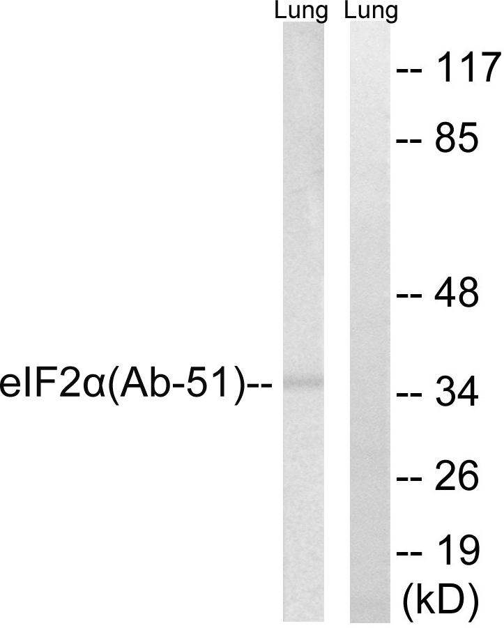
Anti-Phospho-EIF2S1 (S51) Rabbit Monoclonal Antibody
P04387
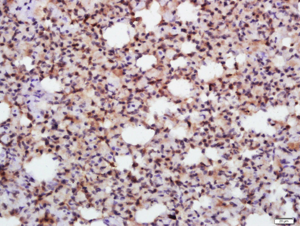
ApplicationsFlow Cytometry, ImmunoFluorescence, Western Blot, ImmunoCytoChemistry, ImmunoHistoChemistry
Product group Antibodies
ReactivityHuman, Mouse, Rat
TargetEIF2S1
Overview
- SupplierBoster Bio
- Product NameAnti-Phospho-EIF2S1 (S51) Rabbit Monoclonal Antibody
- Delivery Days Customer9
- ApplicationsFlow Cytometry, ImmunoFluorescence, Western Blot, ImmunoCytoChemistry, ImmunoHistoChemistry
- CertificationResearch Use Only
- ClonalityMonoclonal
- Clone IDIO-5
- Gene ID1965
- Target nameEIF2S1
- Target descriptioneukaryotic translation initiation factor 2 subunit alpha
- Target synonymsEIF-2, EIF-2A, EIF-2alpha, EIF2, EIF2A, eukaryotic translation initiation factor 2 subunit 1, eIF-2-alpha, eIF2-alpha, eukaryotic translation initiation factor 2, subunit 1 alpha, 35kDa
- HostRabbit
- IsotypeIgG
- Protein IDP05198
- Protein NameEukaryotic translation initiation factor 2 subunit 1
- Scientific DescriptionBoster Bio Anti-Phospho-EIF2S1 (S51) Rabbit Monoclonal Antibody catalog # P04387. Tested in WB, IHC, ICC/IF, Flow Cytometry applications. This antibody reacts with Human, Mouse, Rat.
- ReactivityHuman, Mouse, Rat
- Storage Instruction-20°C
- UNSPSC12352203
References
- Zhao C, Lin M, Pan Y, et al. Blockage of High-Affinity Choline Transporter Increases Visceral Hypersensitivity in Rats with Chronic Stress. Gastroenterol Res Pract. 2018,2018:9252984. doi: 10.1155/2018/9252984Read this paper
Datasheet
MSDS
Related products
Product group Antibodies
Anti-eIF2 alpha AntibodyA95082
ApplicationsWestern Blot, ELISA, ImmunoHistoChemistry
ReactivityHuman, Mouse, Rat
- SizePrice
Product group Antibodies
Anti-EIF2S1 Antibody144-00764
ApplicationsWestern Blot, ImmunoHistoChemistry
ReactivityHuman, Mouse, Rat
TargetEIF2S1
- SizePrice
Product group Antibodies
References
EIF2S1 Polyclonal AntibodyBS-3613R
ApplicationsFlow Cytometry, ImmunoFluorescence, Western Blot, ELISA, ImmunoCytoChemistry, ImmunoHistoChemistry, ImmunoHistoChemistry Frozen, ImmunoHistoChemistry Paraffin
ReactivityBovine, Chicken, Human, Mouse, Rabbit, Rat
TargetEIF2S1
- SizePrice
Product group Antibodies
EIF2S1 AntibodyCSB-PA002295
ApplicationsWestern Blot, ELISA, ImmunoHistoChemistry
ReactivityHuman, Monkey, Mouse, Rat
TargetEIF2S1
- SizePrice
Product group Antibodies
ApplicationsImmunoPrecipitation, Western Blot, ImmunoCytoChemistry, ImmunoHistoChemistry
ReactivityMouse, Rat
TargetEIF2S1
- SizePrice
![eIF2 alpha antibody [N1C3] detects eIF2 alpha protein at cytoplasm by immunofluorescent analysis. Sample: HCT 116 cells were fixed in 4% paraformaldehyde at RT for 15 min. Green: eIF2 alpha protein stained by eIF2 alpha antibody [N1C3] (GTX101241) diluted at 1:500. Blue: Hoechst 33342 staining.](https://www.genetex.com/upload/website/prouct_img/normal/GTX101241/GTX101241_42774_20170628_IFA_w_23060100_284.webp)
Product group Antibodies
eIF2 alpha antibody [N1C3]GTX101241
ApplicationsImmunoFluorescence, Western Blot, ImmunoCytoChemistry
ReactivityHuman, Mouse, Rat
TargetEIF2S1
- SizePrice
Product group Antibodies

EIF2S1 AntibodyLS-C331101
ApplicationsFlow Cytometry, ImmunoFluorescence, ImmunoPrecipitation, Western Blot, ImmunoHistoChemistry
ReactivityHuman, Mouse
TargetEIF2S1
- SizePrice

Product group Antibodies
Anti-EIF2S1 AntibodyHPA064885
ApplicationsWestern Blot, ImmunoCytoChemistry, ImmunoHistoChemistry
ReactivityHuman
TargetEIF2S1
- SizePrice
Product group Antibodies
TargetEIF2S1
- SizePrice
Product group Antibodies
Anti-eIF2alpha AntibodyCAB0764
ApplicationsWestern Blot, ELISA
ReactivityHuman
TargetEIF2S1
- SizePrice