


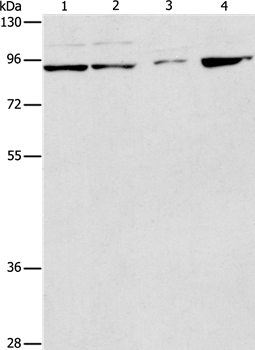
Figure 1. Western blot analysis of PIK3R2 using anti-PIK3R2 antibody (PB9777). Electrophoresis was performed on a 5-20% SDS-PAGE gel at 70V (Stacking gel) / 90V (Resolving gel) for 2-3 hours. The sample well of each lane was loaded with 40 ug of sample under reducing conditions. Lane 1: HELA Whole Cell Lysate, Lane 2: 22RV1 Whole Cell Lysate, Lane 3: MCF-7 Whole Cell Lysate, Lane 4: SW620 Whole Cell Lysate. After electrophoresis, proteins were transferred to a nitrocellulose membrane at 150 mA for 50-90 minutes. Blocked the membrane with 5% non-fat milk/TBS for 1.5 hour at RT. The membrane was incubated with rabbit anti-PIK3R2 antigen affinity purified polyclonal antibody (Catalog # PB9777) at 0.5 microg/mL overnight at 4°C, then washed with TBS-0.1%Tween 3 times with 5 minutes each and probed with a goat anti-rabbit IgG-HRP secondary antibody at a dilution of 1:5000 for 1.5 hour at RT. The signal is developed using an Enhanced Chemiluminescent detection (ECL) kit (Catalog # EK1002) with Tanon 5200 system. A specific band was detected for PIK3R2 at approximately 85 kDa. The expected band size for PIK3R2 is at 85 kDa.
Figure 1. Western blot analysis of PIK3R2 using anti-PIK3R2 antibody (PB9777). Electrophoresis was performed on a 5-20% SDS-PAGE gel at 70V (Stacking gel) / 90V (Resolving gel) for 2-3 hours. The sample well of each lane was loaded with 40 ug of sample under reducing conditions. Lane 1: HELA Whole Cell Lysate, Lane 2: 22RV1 Whole Cell Lysate, Lane 3: MCF-7 Whole Cell Lysate, Lane 4: SW620 Whole Cell Lysate. After electrophoresis, proteins were transferred to a nitrocellulose membrane at 150 mA for 50-90 minutes. Blocked the membrane with 5% non-fat milk/TBS for 1.5 hour at RT. The membrane was incubated with rabbit anti-PIK3R2 antigen affinity purified polyclonal antibody (Catalog # PB9777) at 0.5 microg/mL overnight at 4°C, then washed with TBS-0.1%Tween 3 times with 5 minutes each and probed with a goat anti-rabbit IgG-HRP secondary antibody at a dilution of 1:5000 for 1.5 hour at RT. The signal is developed using an Enhanced Chemiluminescent detection (ECL) kit (Catalog # EK1002) with Tanon 5200 system. A specific band was detected for PIK3R2 at approximately 85 kDa. The expected band size for PIK3R2 is at 85 kDa.
Anti-PI 3 Kinase p85 beta/PIK3R2 Antibody Picoband(r)
PB9777-CARRIER-FREE
ApplicationsWestern Blot
Product group Antibodies
ReactivityBovine, Human
TargetPIK3R2
Overview
- SupplierBoster Bio
- Product NameAnti-PI 3 Kinase p85 beta/PIK3R2 Antibody Picoband(r)
- Delivery Days Customer9
- Application Supplier NoteTested Species: In-house tested species with positive results. Other applications have not been tested. Optimal dilutions should be determined by end users.
- ApplicationsWestern Blot
- CertificationResearch Use Only
- ClonalityPolyclonal
- Concentration500 ug/ml
- Gene ID5296
- Target namePIK3R2
- Target descriptionphosphoinositide-3-kinase regulatory subunit 2
- Target synonymsMPPH, MPPH1, P85B, p85, p85-BETA, p85beta, phosphatidylinositol 3-kinase regulatory subunit beta, PI3-kinase subunit p85-beta, PI3K regulatory subunit beta, phosphatidylinositol 3-kinase 85 kDa regulatory subunit beta, phosphatidylinositol 3-kinase, regulatory subunit, polypeptide 2 (p85 beta), phosphoinositide-3-kinase regulatory subunit beta, phosphoinositide-3-kinase, regulatory subunit 2 (beta), ptdIns-3-kinase regulatory subunit p85-beta
- HostRabbit
- IsotypeIgG
- Protein IDO00459
- Protein NamePhosphatidylinositol 3-kinase regulatory subunit beta
- Scientific DescriptionBoster Bio Anti-PI 3 Kinase p85 beta/PIK3R2 Antibody Picoband® catalog # PB9777. Tested in WB applications. This antibody reacts with Human. The brand Picoband indicates this is a premium antibody that guarantees superior quality, high affinity, and strong signals with minimal background in Western blot applications. Only our best-performing antibodies are designated as Picoband, ensuring unmatched performance.
- ReactivityBovine, Human
- Storage Instruction-20°C,2°C to 8°C
- UNSPSC12352203
Related products
Product group Antibodies
Anti-PIK3R2 AntibodyA37283
ApplicationsWestern Blot, ImmunoHistoChemistry
ReactivityHuman, Rat
- SizePrice
Product group Antibodies
Anti-PIK3R2 Antibody144-64925
ApplicationsImmunoFluorescence, Western Blot, ImmunoHistoChemistry
ReactivityHuman, Mouse, Rat
TargetPIK3R2
- SizePrice
Product group Antibodies
ApplicationsImmunoFluorescence, Western Blot, ELISA, ImmunoCytoChemistry, ImmunoHistoChemistry, ImmunoHistoChemistry Frozen, ImmunoHistoChemistry Paraffin
ReactivityBovine, Canine, Equine, Human, Mouse, Rabbit, Rat
TargetPIK3R2
- SizePrice
Product group Antibodies
PIK3R2 AntibodyCSB-PA003770
ApplicationsWestern Blot, ELISA
ReactivityHuman, Mouse, Rat
TargetPIK3R2
- SizePrice
Product group Antibodies
PIK3R2 Polyclonal AntibodyCAC14862
ApplicationsWestern Blot, ELISA, ImmunoHistoChemistry
TargetPIK3R2
- SizePrice
![WB analysis of various cell lysates using PI3K p85 beta antibody [8D9-D5-F8] at a dilution of 1:1000.](https://www.genetex.com/upload/website/prouct_img/normal/GTX16481/GTX16481_WB_w_23060620_448.webp)
Product group Antibodies
ApplicationsImmunoDiffusion, Western Blot
ReactivityHuman, Mouse, Rat
TargetPIK3R2
- SizePrice

Product group Antibodies
Anti-PIK3R2 AntibodyHPA069291
ApplicationsWestern Blot, ImmunoCytoChemistry, ImmunoHistoChemistry
ReactivityHuman
TargetPIK3R2
- SizePrice
Product group Antibodies
ApplicationsWestern Blot, ELISA
ReactivityHuman
TargetPIK3R2
- SizePrice
Product group Antibodies

PIK3R2 / p85 Beta AntibodyLS-C406166
ApplicationsWestern Blot, ELISA, ImmunoHistoChemistry
ReactivityHuman, Mouse, Rat
TargetPIK3R2
- SizePrice