


Figure 1. Western blot analysis of PIAS2 using anti-PIAS2 antibody (A04130-3). Electrophoresis was performed on a 5-20% SDS-PAGE gel at 70V (Stacking gel) / 90V (Resolving gel) for 2-3 hours. The sample well of each lane was loaded with 50ug of sample under reducing conditions. Lane 1: human K562 whole cell lysates, Lane 2: human Hela whole cell lysates, Lane 3: human HepG2 whole cell lysates, Lane 4: human HEK293 whole cell lysates, Lane 5: human HL-60 whole cell lysates, Lane 6: human Raji whole cell lysates, Lane 7: human A431 whole cell lysates, Lane 8: rat brain tissue lysates, Lane 9: rat C6 whole cell lysates, Lane 10: mouse brain tissue lysates, Lane 11: mouse testis tissue lysates, Lane 12: mouse Neuro-2a whole cell lysates, Lane 13: mouse RAW264.7 whole cell lysates. After Electrophoresis, proteins were transferred to a Nitrocellulose membrane at 150mA for 50-90 minutes. Blocked the membrane with 5% Non-fat Milk/ TBS for 1.5 hour at RT. The membrane was incubated with rabbit anti-PIAS2 antigen affinity purified polyclonal antibody (Catalog # A04130-3) at 0.25 microg/mL overnight at 4°C, then washed with TBS-0.1%Tween 3 times with 5 minutes each and probed with a goat anti-rabbit IgG-HRP secondary antibody at a dilution of 1:5000 for 1.5 hour at RT. The signal is developed using an Enhanced Chemiluminescent detection (ECL) kit (Catalog # EK1002) with Tanon 5200 system. A specific band was detected for PIAS2 at approximately 68KD. The expected band size for PIAS2 is at 68KD.
. Overlay histogram showing U937 cells stained with A04130-3 (Blue line). To facilitate intracellular staining, cells were fixed with 4% paraformaldehyde and permeabilized with permeabilization buffer. The cells were blocked with 10% normal goat serum. And then incubated with rabbit anti-PIAS2 Antibody (A04130-3, 1microg/1x106 cells) for 30 min at 20°C. DyLight®488 conjugated goat anti-rabbit IgG (BA1127, 5-10microg/1x106 cells) was used as secondary antibody for 30 minutes at 20°C. Isotype control antibody (Green line) was rabbit IgG (1microg/1x106) used under the same conditions. Unlabelled sample without incubation with primary antibody and secondary antibody (Red line) was used as a blank control.")
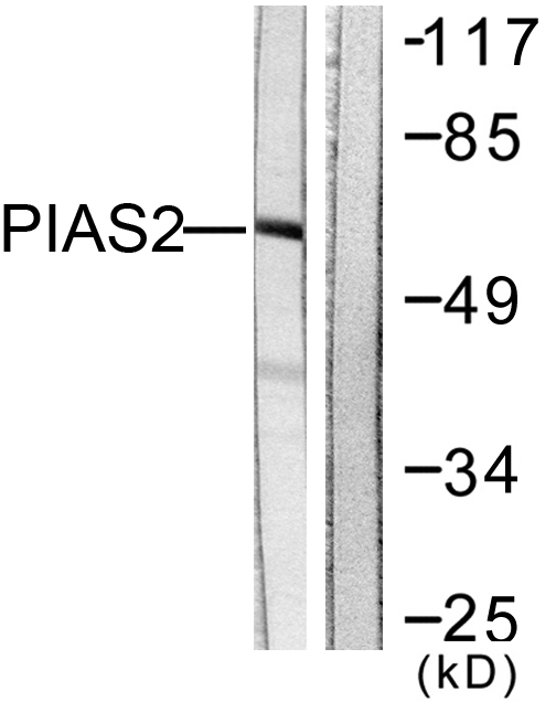
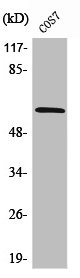
Figure 1. Western blot analysis of PIAS2 using anti-PIAS2 antibody (A04130-3). Electrophoresis was performed on a 5-20% SDS-PAGE gel at 70V (Stacking gel) / 90V (Resolving gel) for 2-3 hours. The sample well of each lane was loaded with 50ug of sample under reducing conditions. Lane 1: human K562 whole cell lysates, Lane 2: human Hela whole cell lysates, Lane 3: human HepG2 whole cell lysates, Lane 4: human HEK293 whole cell lysates, Lane 5: human HL-60 whole cell lysates, Lane 6: human Raji whole cell lysates, Lane 7: human A431 whole cell lysates, Lane 8: rat brain tissue lysates, Lane 9: rat C6 whole cell lysates, Lane 10: mouse brain tissue lysates, Lane 11: mouse testis tissue lysates, Lane 12: mouse Neuro-2a whole cell lysates, Lane 13: mouse RAW264.7 whole cell lysates. After Electrophoresis, proteins were transferred to a Nitrocellulose membrane at 150mA for 50-90 minutes. Blocked the membrane with 5% Non-fat Milk/ TBS for 1.5 hour at RT. The membrane was incubated with rabbit anti-PIAS2 antigen affinity purified polyclonal antibody (Catalog # A04130-3) at 0.25 microg/mL overnight at 4°C, then washed with TBS-0.1%Tween 3 times with 5 minutes each and probed with a goat anti-rabbit IgG-HRP secondary antibody at a dilution of 1:5000 for 1.5 hour at RT. The signal is developed using an Enhanced Chemiluminescent detection (ECL) kit (Catalog # EK1002) with Tanon 5200 system. A specific band was detected for PIAS2 at approximately 68KD. The expected band size for PIAS2 is at 68KD.
Anti-PIAS2 Antibody Picoband(r)
A04130-3-CARRIER-FREE
ApplicationsFlow Cytometry, Western Blot, ELISA
Product group Antibodies
ReactivityHuman, Mouse, Rat
TargetPIAS2
Overview
- SupplierBoster Bio
- Product NameAnti-PIAS2 Antibody Picoband(r)
- Delivery Days Customer9
- ApplicationsFlow Cytometry, Western Blot, ELISA
- CertificationResearch Use Only
- ClonalityPolyclonal
- Concentration500 ug/ml
- Gene ID9063
- Target namePIAS2
- Target descriptionprotein inhibitor of activated STAT 2
- Target synonymsARIP3, DIP, MIZ1, PIASX, SIZ2, ZMIZ4, E3 SUMO-protein ligase PIAS2, DAB2-interacting protein, E3 SUMO-protein transferase PIAS2, androgen receptor-interacting protein 3, msx-interacting zinc finger protein, protein inhibitor of activated STAT X, zinc finger, MIZ-type containing 4
- HostRabbit
- IsotypeIgG
- Protein IDO75928
- Protein NameE3 SUMO-protein ligase PIAS2
- Scientific DescriptionBoster Bio Anti-PIAS2 Antibody Picoband® catalog # A04130-3. Tested in ELISA, Flow Cytometry, WB applications. This antibody reacts with Human, Mouse, Rat. The brand Picoband indicates this is a premium antibody that guarantees superior quality, high affinity, and strong signals with minimal background in Western blot applications. Only our best-performing antibodies are designated as Picoband, ensuring unmatched performance.
- ReactivityHuman, Mouse, Rat
- Storage Instruction-20°C,2°C to 8°C
- UNSPSC12352203
Related products
Product group Antibodies
Anti-PIAS2 AntibodyA97347
ApplicationsWestern Blot, ELISA
ReactivityHuman, Mouse, Rat
- SizePrice
Product group Antibodies
Anti-PIAS2 Antibody144-05654
ApplicationsWestern Blot
ReactivityHuman, Mouse
TargetPIAS2
- SizePrice
Product group Antibodies
PIAS2 Recombinant AntibodyBSM-62274R
ApplicationsImmunoFluorescence, Western Blot, ImmunoCytoChemistry
ReactivityHuman, Mouse, Rat
TargetPIAS2
- SizePrice
Product group Antibodies
PIAS2 AntibodyCSB-PA003781
ApplicationsWestern Blot, ELISA
ReactivityHuman, Monkey, Mouse, Rat
TargetPIAS2
- SizePrice
Product group Antibodies

Goat anti-PIAS2EB07176
ApplicationsELISA, ImmunoHistoChemistry
ReactivityCanine, Human, Mouse, Rat
TargetPIAS2
- SizePrice
Product group Antibodies

PIAS2 / PIASX AntibodyLS-C402922
ApplicationsELISA, ImmunoHistoChemistry
ReactivityHuman, Mouse
TargetPIAS2
- SizePrice
Product group Antibodies
PIAS2 antibody [N1N3]GTX103275
ApplicationsWestern Blot
ReactivityHuman
TargetPIAS2
- SizePrice

Product group Antibodies
Anti-PIAS2 AntibodyHPA068792
ApplicationsImmunoCytoChemistry, ImmunoHistoChemistry
ReactivityHuman
TargetPIAS2
- SizePrice
Product group Antibodies
ApplicationsWestern Blot, ELISA
ReactivityMouse
TargetPIAS2
- SizePrice