


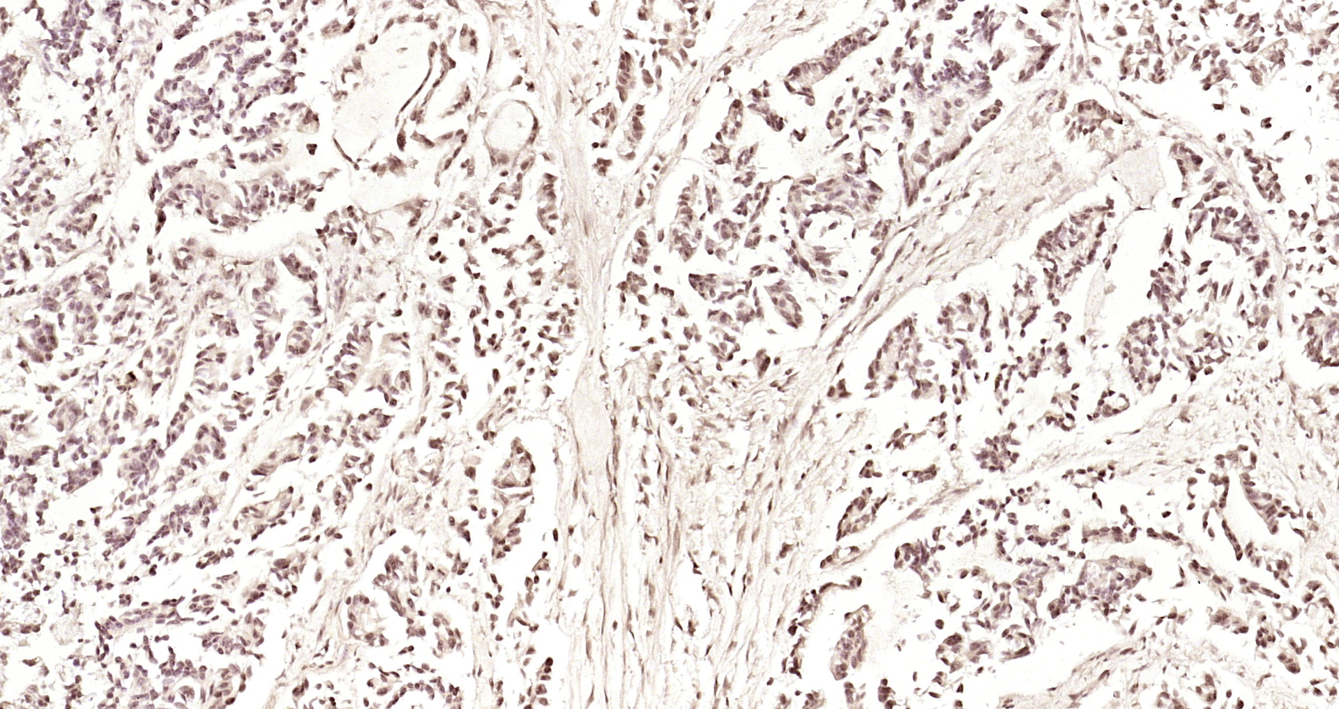
Immunofluorescent staining of human cell line U-2 OS shows localization to cytosol.
Immunofluorescent staining of human cell line U-2 OS shows localization to cytosol.
Anti-PIK3R5 Antibody
HPA044505
ApplicationsImmunoCytoChemistry
Product group Antibodies
ReactivityHuman
TargetPIK3R5
Overview
- SupplierAtlas Antibodies
- Product NameAnti-PIK3R5 Antibody
- Delivery Days Customer4
- ApplicationsImmunoCytoChemistry
- CertificationResearch Use Only
- ClonalityPolyclonal
- ConjugateUnconjugated
- Gene ID23533
- Target namePIK3R5
- Target descriptionphosphoinositide-3-kinase regulatory subunit 5
- Target synonymsF730038I15Rik, FOAP-2, P101-PI3K, p101, phosphoinositide 3-kinase regulatory subunit 5, PI3-kinase p101 subunit, phosphatidylinositol 4,5-bisphosphate 3-kinase regulatory subunit, protein FOAP-2, ptdIns-3-kinase p101
- HostRabbit
- IsotypeIgG
- Protein IDQ8WYR1
- Protein NamePhosphoinositide 3-kinase regulatory subunit 5
- Scientific DescriptionRecombinant Protein Epitope Signature Tag (PrEST) antigen sequence
- ReactivityHuman
- Storage Instruction-20°C,2°C to 8°C
- UNSPSC41116161
Datasheet
MSDS
Related products
Product group Antibodies
PIK3R5 AntibodyCSB-PA008386
ApplicationsWestern Blot, ELISA, ImmunoHistoChemistry
ReactivityHuman, Mouse
TargetPIK3R5
- SizePrice

Product group Antibodies
Anti-PIK3R5 AntibodyA98628
ApplicationsELISA, ImmunoHistoChemistry
ReactivityHuman, Mouse
- SizePrice

Product group Antibodies
Anti-PIK3R5 Antibody Picoband(r)A02827-2-CARRIER-FREE
ApplicationsFlow Cytometry, Western Blot, ELISA
ReactivityHuman, Mouse, Rat
TargetPIK3R5
- SizePrice
Product group Antibodies

PIK3R5 AntibodyLS-C760976
ApplicationsWestern Blot, ImmunoHistoChemistry
ReactivityChicken, Human, Mouse, Porcine, Rat
TargetPIK3R5
- SizePrice

Product group Antibodies
Anti-PIK3R5 AntibodyHPA052247
ApplicationsImmunoHistoChemistry
ReactivityHuman
TargetPIK3R5
- SizePrice

Product group Antibodies
Anti-PIK3R5 AntibodyHPA052412
ApplicationsWestern Blot, ImmunoCytoChemistry
ReactivityHuman
TargetPIK3R5
- SizePrice
Product group Antibodies
PIK3R5 Polyclonal AntibodyBS-7467R
ApplicationsImmunoFluorescence, ELISA, ImmunoCytoChemistry, ImmunoHistoChemistry, ImmunoHistoChemistry Frozen, ImmunoHistoChemistry Paraffin
ReactivityBovine, Equine, Human, Mouse, Porcine, Rabbit, Rat, Sheep
TargetPIK3R5
- SizePrice
Product group Antibodies
ApplicationsImmunoHistoChemistry, ImmunoHistoChemistry Paraffin
ReactivityHuman, Rat
TargetPIK3R5
- SizePrice