


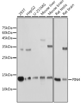
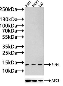
Figure 1. Western blot analysis of PIN4 using anti-PIN4 antibody (M05181-1). Electrophoresis was performed on a 5-20% SDS-PAGE gel at 70V (Stacking gel) / 90V (Resolving gel) for 2-3 hours. The sample well of each lane was loaded with 30 ug of sample under reducing conditions. Lane 1: human HepG2 whole cell lysates, Lane 2: human Hela whole cell lysates, Lane 3: rat liver tissue lysates, Lane 4: rat brain tissue lysates. Lane 5: mouse liver tissue lysates. Lane 6: mouse brain tissue lysates. After electrophoresis, proteins were transferred to a nitrocellulose membrane at 150 mA for 50-90 minutes. Blocked the membrane with 5% non-fat milk/TBS for 1.5 hour at RT. The membrane was incubated with rabbit anti-PIN4 antigen affinity purified monoclonal antibody (Catalog # M05181-1) at 1:500 overnight at 4°C, then washed with TBS-0.1%Tween 3 times with 5 minutes each and probed with a goat anti-rabbit IgG-HRP secondary antibody at a dilution of 1:500 for 1.5 hour at RT. The signal is developed using an Enhanced Chemiluminescent detection (ECL) kit (Catalog # EK1002) with Tanon 5200 system. A specific band was detected for PIN4 at approximately 14 kDa. The expected band size for PIN4 is at 14 kDa.
. PIN4 was detected in a paraffin-embedded section of human prostate cancer tissue. Heat mediated antigen retrieval was performed in EDTA buffer (pH 8.0, epitope retrieval solution). The tissue section was blocked with 10% goat serum. The tissue section was then incubated with 1:50 rabbit anti-PIN4 Antibody (M05181-1) overnight at 4°C. Peroxidase Conjugated Goat Anti-rabbit IgG was used as secondary antibody and incubated for 30 minutes at 37°C. The tissue section was developed using HRP Conjugated Rabbit IgG Super Vision Assay Kit (Catalog # SV0002) with DAB as the chromogen.")
. PIN4 was detected in a paraffin-embedded section of human prostate cancer tissue. Heat mediated antigen retrieval was performed in EDTA buffer (pH 8.0, epitope retrieval solution). The tissue section was blocked with 10% goat serum. The tissue section was then incubated with 1:50 rabbit anti-PIN4 Antibody (M05181-1) overnight at 4°C. Peroxidase Conjugated Goat Anti-rabbit IgG was used as secondary antibody and incubated for 30 minutes at 37°C. The tissue section was developed using HRP Conjugated Rabbit IgG Super Vision Assay Kit (Catalog # SV0002) with DAB as the chromogen.")
Figure 1. Western blot analysis of PIN4 using anti-PIN4 antibody (M05181-1). Electrophoresis was performed on a 5-20% SDS-PAGE gel at 70V (Stacking gel) / 90V (Resolving gel) for 2-3 hours. The sample well of each lane was loaded with 30 ug of sample under reducing conditions. Lane 1: human HepG2 whole cell lysates, Lane 2: human Hela whole cell lysates, Lane 3: rat liver tissue lysates, Lane 4: rat brain tissue lysates. Lane 5: mouse liver tissue lysates. Lane 6: mouse brain tissue lysates. After electrophoresis, proteins were transferred to a nitrocellulose membrane at 150 mA for 50-90 minutes. Blocked the membrane with 5% non-fat milk/TBS for 1.5 hour at RT. The membrane was incubated with rabbit anti-PIN4 antigen affinity purified monoclonal antibody (Catalog # M05181-1) at 1:500 overnight at 4°C, then washed with TBS-0.1%Tween 3 times with 5 minutes each and probed with a goat anti-rabbit IgG-HRP secondary antibody at a dilution of 1:500 for 1.5 hour at RT. The signal is developed using an Enhanced Chemiluminescent detection (ECL) kit (Catalog # EK1002) with Tanon 5200 system. A specific band was detected for PIN4 at approximately 14 kDa. The expected band size for PIN4 is at 14 kDa.
Anti-PIN4 Rabbit Monoclonal Antibody
M05181-1
ApplicationsImmunoFluorescence, Western Blot, ImmunoCytoChemistry, ImmunoHistoChemistry
Product group Antibodies
ReactivityHuman, Mouse, Rat
TargetPIN4
Overview
- SupplierBoster Bio
- Product NameAnti-PIN4 Rabbit Monoclonal Antibody
- Delivery Days Customer9
- ApplicationsImmunoFluorescence, Western Blot, ImmunoCytoChemistry, ImmunoHistoChemistry
- CertificationResearch Use Only
- ClonalityMonoclonal
- Clone ID29P19
- Gene ID5303
- Target namePIN4
- Target descriptionpeptidylprolyl cis/trans isomerase, NIMA-interacting 4
- Target synonymsEPVH, PAR14, PAR17, hEPVH, hPar14, hPar17, peptidyl-prolyl cis-trans isomerase NIMA-interacting 4, PPIase PIN4, eukaryotic parvulin homolog, parvulin, parvulin-14, parvulin-17, peptidyl-prolyl cis-trans isomerase Pin4, peptidyl-prolyl cis/trans isomerase EPVH, protein (peptidylprolyl cis/trans isomerase) NIMA-interacting, 4 (parvulin), rotamase PIN4
- HostRabbit
- IsotypeIgG
- Protein IDQ9Y237
- Protein NamePeptidyl-prolyl cis-trans isomerase NIMA-interacting 4
- Scientific DescriptionBoster Bio Anti-PIN4 Rabbit Monoclonal Antibody catalog # M05181-1. Tested in WB, IHC, ICC/IF applications. This antibody reacts with Human, Mouse, Rat.
- ReactivityHuman, Mouse, Rat
- Storage Instruction-20°C
- UNSPSC12352203
Related products
Product group Antibodies
Anti-PIN4 AntibodyA307628
ApplicationsWestern Blot
ReactivityHuman, Mouse, Rat
- SizePrice

Product group Antibodies
PIN4 Recombinant AntibodyBSM-62647R
ApplicationsImmunoFluorescence, Western Blot, ImmunoCytoChemistry, ImmunoHistoChemistry, ImmunoHistoChemistry Frozen, ImmunoHistoChemistry Paraffin
ReactivityHuman, Mouse, Rat
TargetPIN4
- SizePrice
Product group Antibodies
PIN4 AntibodyCSB-PA861478LA01HU
ApplicationsImmunoFluorescence, Western Blot, ELISA, ImmunoHistoChemistry
ReactivityHuman, Mouse, Rat
TargetPIN4
- SizePrice
Product group Antibodies
Pin4 Polyclonal AntibodyCAC11415
ApplicationsImmunoFluorescence, Western Blot, ELISA, ImmunoHistoChemistry
ReactivityMouse, Rat
TargetPIN4
- SizePrice

Product group Antibodies
Anti-PIN4 AntibodyHPA054483
ApplicationsImmunoCytoChemistry, ImmunoHistoChemistry
ReactivityHuman
TargetPIN4
- SizePrice
Product group Antibodies
PIN4 antibody, InternalGTX45041
ApplicationsWestern Blot
ReactivityHuman
TargetPIN4
- SizePrice
Product group Antibodies
Anti-PIN4 AntibodyCAB18173
ApplicationsWestern Blot, ELISA
ReactivityHuman
TargetPIN4
- SizePrice