


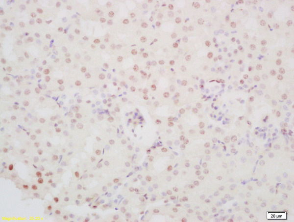
Immunohistochemical staining of human testis shows strong nuclear positivity in cells in seminiferous ducts.

Immunohistochemical staining of human testis shows strong nuclear positivity in cells in seminiferous ducts.
Anti-PINX1 Antibody
HPA023139
ApplicationsWestern Blot, ImmunoHistoChemistry
Product group Antibodies
ReactivityHuman
TargetPINX1
Overview
- SupplierAtlas Antibodies
- Product NameAnti-PINX1 Antibody
- Delivery Days Customer4
- ApplicationsWestern Blot, ImmunoHistoChemistry
- CertificationResearch Use Only
- ClonalityPolyclonal
- ConjugateUnconjugated
- Gene ID54984
- Target namePINX1
- Target descriptionPIN2 (TERF1) interacting telomerase inhibitor 1
- Target synonymsGno1, LPTL, LPTS, Pxr1, PIN2/TERF1-interacting telomerase inhibitor 1, 67-11-3 protein, PIN2-interacting protein 1, TRF1-interacting protein 1, hepatocellular carcinoma-related putative tumor suppressor, liver-related putative tumor suppressor, pin2-interacting protein X1, protein 67-11-3
- HostRabbit
- IsotypeIgG
- Protein IDQ96BK5
- Protein NamePIN2/TERF1-interacting telomerase inhibitor 1
- Scientific DescriptionRecombinant Protein Epitope Signature Tag (PrEST) antigen sequence
- ReactivityHuman
- Storage Instruction-20°C,2°C to 8°C
- UNSPSC41116161
Datasheet
MSDS
Related products
Product group Antibodies
Anti-PINX1 AntibodyA100650
ApplicationsELISA, ImmunoHistoChemistry
ReactivityHuman
- SizePrice
Product group Antibodies
PINX-1 Polyclonal AntibodyBS-9594R
ApplicationsFlow Cytometry, ImmunoFluorescence, ImmunoHistoChemistry, ImmunoHistoChemistry Frozen, ImmunoHistoChemistry Paraffin
ReactivityHuman, Mouse, Rat
TargetPINX1
- SizePrice
Product group Antibodies

Goat anti-PINX1EB05324
ApplicationsWestern Blot, ELISA
ReactivityBovine, Canine, Human, Mouse, Porcine, Rat
TargetPINX1
- SizePrice
Product group Antibodies
PINX1 Polyclonal AntibodyCAC14666
ApplicationsWestern Blot, ELISA
ReactivityMouse
TargetPINX1
- SizePrice
Product group Antibodies
PINX1 AntibodyCSB-PA030159
ApplicationsELISA, ImmunoHistoChemistry
ReactivityHuman
TargetPINX1
- SizePrice
Product group Antibodies

PINX1 AntibodyLS-C501513
ApplicationsWestern Blot, ELISA
ReactivityHuman, Mouse
TargetPINX1
- SizePrice

Product group Antibodies
Anti-PINX1 AntibodyHPA023146
ApplicationsImmunoCytoChemistry
ReactivityHuman
TargetPINX1
- SizePrice