


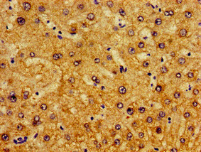
Immunohistochemical staining of human skin shows strong cytoplasmic and membranous positivity in squamous epithelial cells.

Immunohistochemical staining of human skin shows strong cytoplasmic and membranous positivity in squamous epithelial cells.
Anti-PIR Antibody
HPA000697
ApplicationsWestern Blot
Product group Antibodies
ReactivityHuman, Rat
TargetPIR
Overview
- SupplierAtlas Antibodies
- Product NameAnti-PIR Antibody
- Delivery Days Customer4
- ApplicationsWestern Blot
- CertificationResearch Use Only
- ClonalityPolyclonal
- ConjugateUnconjugated
- Gene ID8544
- Target namePIR
- Target descriptionpirin
- Target synonymspirin, pirin (iron-binding nuclear protein), probable quercetin 2,3-dioxygenase PIR, probable quercetinase
- HostRabbit
- IsotypeIgG
- Protein IDO00625
- Protein NamePirin
- Scientific DescriptionRecombinant Protein Epitope Signature Tag (PrEST) antigen sequence
- ReactivityHuman, Rat
- Storage Instruction-20°C,2°C to 8°C
- UNSPSC41116161
Datasheet
MSDS
Related products
Product group Antibodies
PIR AntibodyCSB-PA018032LA01HU
ApplicationsImmunoFluorescence, ELISA, ImmunoHistoChemistry
ReactivityHuman
TargetPIR
- SizePrice

Product group Antibodies
Anti-PIR Antibody Picoband(r)A03468-1-CARRIER-FREE
ApplicationsFlow Cytometry, ImmunoFluorescence, Western Blot, ELISA, ImmunoCytoChemistry
ReactivityHuman, Monkey, Mouse, Rat
TargetPIR
- SizePrice
Product group Antibodies

Goat anti-PirinEB06437
ApplicationsWestern Blot, ELISA, ImmunoHistoChemistry
ReactivityHuman, Mouse
TargetPIR
- SizePrice
Product group Antibodies

Pirin / PIR AntibodyLS-C672320
ApplicationsELISA, ImmunoHistoChemistry, ImmunoHistoChemistry Paraffin
ReactivityHuman
TargetPIR
- SizePrice
Product group Antibodies
Pirin antibodyGTX113584
ApplicationsImmunoFluorescence, Western Blot, ImmunoCytoChemistry, ImmunoHistoChemistry, ImmunoHistoChemistry Paraffin
ReactivityHuman, Mouse, Rat
TargetPIR
- SizePrice
Product group Antibodies
PIR Polyclonal AntibodyCAC13159
ApplicationsImmunoFluorescence, ELISA, ImmunoHistoChemistry
TargetPIR
- SizePrice