


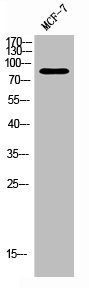
Figure 1. Western blot analysis of PKC Alpha/PRKCA using anti-PKC Alpha/PRKCA antibody (A00743-1). Electrophoresis was performed on a 5-20% SDS-PAGE gel at 70V (Stacking gel) / 90V (Resolving gel) for 2-3 hours. The sample well of each lane was loaded with 30 ug of sample under reducing conditions. Lane 1: human Jurkat whole cell lysates, Lane 2: human 293T cell lysates, Lane 3: human Hela whole cell lysates, Lane 4: human K562 whole cell lysates, Lane 5: rat C6 whole cell lysates, Lane 6: mouse NIH/3T3 whole cell lysates. After electrophoresis, proteins were transferred to a nitrocellulose membrane at 150 mA for 50-90 minutes. Blocked the membrane with 5% non-fat milk/TBS for 1.5 hour at RT. The membrane was incubated with rabbit anti-PKC Alpha/PRKCA antigen affinity purified polyclonal antibody (Catalog # A00743-1) at 0.5 microg/mL overnight at 4°C, then washed with TBS-0.1%Tween 3 times with 5 minutes each and probed with a goat anti-rabbit IgG-HRP secondary antibody at a dilution of 1:5000 for 1.5 hour at RT. The signal is developed using an Enhanced Chemiluminescent detection (ECL) kit (Catalog # EK1002) with Tanon 5200 system. A specific band was detected for PKC Alpha/PRKCA at approximately 80 kDa. The expected band size for PKC Alpha/PRKCA is at 77 kDa.
. Overlay histogram showing SiHa cells stained with A00743-1 (Blue line). To facilitate intracellular staining, cells were fixed with 4% paraformaldehyde and permeabilized with permeabilization buffer. The cells were blocked with 10% normal goat serum. And then incubated with rabbit anti-PKC Alpha/PRKCA Antibody (A00743-1, 1 microg/1x106 cells) for 30 min at 20°C. DyLight®488 conjugated goat anti-rabbit IgG (BA1127, 5-10 microg/1x106 cells) was used as secondary antibody for 30 minutes at 20°C. Isotype control antibody (Green line) was rabbit IgG (1 microg/1x106) used under the same conditions. Unlabelled sample (Red line) was also used as a control.")
Figure 1. Western blot analysis of PKC Alpha/PRKCA using anti-PKC Alpha/PRKCA antibody (A00743-1). Electrophoresis was performed on a 5-20% SDS-PAGE gel at 70V (Stacking gel) / 90V (Resolving gel) for 2-3 hours. The sample well of each lane was loaded with 30 ug of sample under reducing conditions. Lane 1: human Jurkat whole cell lysates, Lane 2: human 293T cell lysates, Lane 3: human Hela whole cell lysates, Lane 4: human K562 whole cell lysates, Lane 5: rat C6 whole cell lysates, Lane 6: mouse NIH/3T3 whole cell lysates. After electrophoresis, proteins were transferred to a nitrocellulose membrane at 150 mA for 50-90 minutes. Blocked the membrane with 5% non-fat milk/TBS for 1.5 hour at RT. The membrane was incubated with rabbit anti-PKC Alpha/PRKCA antigen affinity purified polyclonal antibody (Catalog # A00743-1) at 0.5 microg/mL overnight at 4°C, then washed with TBS-0.1%Tween 3 times with 5 minutes each and probed with a goat anti-rabbit IgG-HRP secondary antibody at a dilution of 1:5000 for 1.5 hour at RT. The signal is developed using an Enhanced Chemiluminescent detection (ECL) kit (Catalog # EK1002) with Tanon 5200 system. A specific band was detected for PKC Alpha/PRKCA at approximately 80 kDa. The expected band size for PKC Alpha/PRKCA is at 77 kDa.
Anti-PKC Alpha/PRKCA Antibody Picoband(r)
A00743-1-CARRIER-FREE
ApplicationsFlow Cytometry, Western Blot, ELISA
Product group Antibodies
ReactivityHuman, Mouse, Rat
TargetPRKCA
Overview
- SupplierBoster Bio
- Product NameAnti-PKC Alpha/PRKCA Antibody Picoband(r)
- Delivery Days Customer9
- ApplicationsFlow Cytometry, Western Blot, ELISA
- CertificationResearch Use Only
- ClonalityPolyclonal
- Concentration500 ug/ml
- Gene ID5578
- Target namePRKCA
- Target descriptionprotein kinase C alpha
- Target synonymsAAG6, PKC-alpha, PKCA, PKCI+/-, PKCalpha, PRKACA, protein kinase C alpha type, PKC-A, aging-associated gene 6
- HostRabbit
- IsotypeIgG
- Protein IDP17252
- Protein NameProtein kinase C alpha type
- Scientific DescriptionBoster Bio Anti-PKC Alpha/PRKCA Antibody Picoband® catalog # A00743-1. Tested in ELISA, Flow Cytometry, WB applications. This antibody reacts with Human, Mouse, Rat. The brand Picoband indicates this is a premium antibody that guarantees superior quality, high affinity, and strong signals with minimal background in Western blot applications. Only our best-performing antibodies are designated as Picoband, ensuring unmatched performance.
- ReactivityHuman, Mouse, Rat
- Storage Instruction-20°C,2°C to 8°C
- UNSPSC12352203
Related products
Product group Antibodies
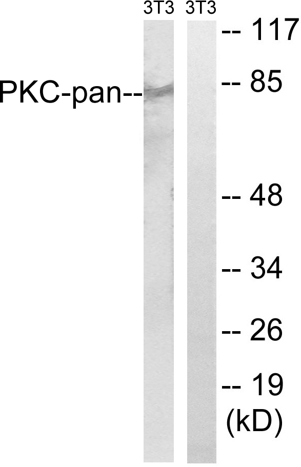
Anti-PKC-pan AntibodyA94967
ApplicationsImmunoFluorescence, Western Blot, ELISA, ImmunoHistoChemistry
ReactivityHuman, Mouse, Rat
- SizePrice
Product group Antibodies

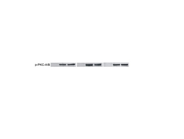
PKC alpha (Phospho-Tyr657) AntibodyABX012471
ApplicationsWestern Blot, ELISA
- SizePrice
Product group Antibodies
Anti-PRKCA Antibody144-00267
ApplicationsImmunoFluorescence, Western Blot, ImmunoHistoChemistry
ReactivityHuman
TargetPRKCA
- SizePrice
Product group Antibodies
References
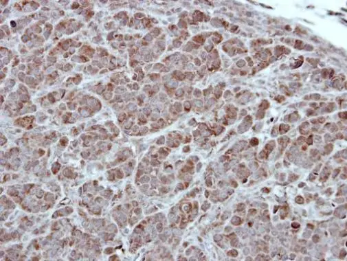
ApplicationsImmunoFluorescence, Western Blot, ELISA, ImmunoCytoChemistry, ImmunoHistoChemistry, ImmunoHistoChemistry Frozen, ImmunoHistoChemistry Paraffin
ReactivityBovine, Canine, Chicken, Equine, Human, Mouse, Rat
TargetPRKCA
- SizePrice
Product group Antibodies
ApplicationsImmunoFluorescence, Western Blot, ELISA, ImmunoHistoChemistry
ReactivityHuman, Mouse, Rat
TargetPRKCA
- SizePrice
Product group Antibodies
Prkca Polyclonal AntibodyCAC09189
ApplicationsImmunoFluorescence, ELISA, ImmunoHistoChemistry
TargetPRKCA
- SizePrice
Product group Antibodies

PRKCA / PKC-Alpha AntibodyLS-C402927
ApplicationsWestern Blot, ELISA
ReactivityHuman, Mouse, Rat
TargetPRKCA
- SizePrice

Product group Antibodies
Anti-PRKCA AntibodyHPA006564
ApplicationsWestern Blot, ImmunoCytoChemistry, ImmunoHistoChemistry
ReactivityHuman, Mouse, Rat
TargetPRKCA
- SizePrice
Product group Antibodies
PKC alpha antibody [C1C3]GTX111096
ApplicationsWestern Blot, ImmunoHistoChemistry, ImmunoHistoChemistry Paraffin
ReactivityHuman, Mouse
TargetPRKCA
- SizePrice
Product group Antibodies
TargetPRKCA
- SizePrice