


Figure 1. Western blot analysis of PLA2G6 using anti-PLA2G6 antibody (A01648-1). Electrophoresis was performed on a 5-20% SDS-PAGE gel at 70V (Stacking gel) / 90V (Resolving gel) for 2-3 hours. The sample well of each lane was loaded with 50ug of sample under reducing conditions. Lane 1: human Hela whole cell lysates. After Electrophoresis, proteins were transferred to a Nitrocellulose membrane at 150mA for 50-90 minutes. Blocked the membrane with 5% Non-fat Milk/ TBS for 1.5 hour at RT. The membrane was incubated with rabbit anti-PLA2G6 antigen affinity purified polyclonal antibody (Catalog # A01648-1) at 0.5 microg/mL overnight at 4°C, then washed with TBS-0.1%Tween 3 times with 5 minutes each and probed with a goat anti-rabbit IgG-HRP secondary antibody at a dilution of 1:10000 for 1.5 hour at RT. The signal is developed using an Enhanced Chemiluminescent detection (ECL) kit (Catalog # EK1002) with Tanon 5200 system. A specific band was detected for PLA2G6 at approximately 90KD. The expected band size for PLA2G6 is at 90KD.
Figure 1. Western blot analysis of PLA2G6 using anti-PLA2G6 antibody (A01648-1). Electrophoresis was performed on a 5-20% SDS-PAGE gel at 70V (Stacking gel) / 90V (Resolving gel) for 2-3 hours. The sample well of each lane was loaded with 50ug of sample under reducing conditions. Lane 1: human Hela whole cell lysates. After Electrophoresis, proteins were transferred to a Nitrocellulose membrane at 150mA for 50-90 minutes. Blocked the membrane with 5% Non-fat Milk/ TBS for 1.5 hour at RT. The membrane was incubated with rabbit anti-PLA2G6 antigen affinity purified polyclonal antibody (Catalog # A01648-1) at 0.5 microg/mL overnight at 4°C, then washed with TBS-0.1%Tween 3 times with 5 minutes each and probed with a goat anti-rabbit IgG-HRP secondary antibody at a dilution of 1:10000 for 1.5 hour at RT. The signal is developed using an Enhanced Chemiluminescent detection (ECL) kit (Catalog # EK1002) with Tanon 5200 system. A specific band was detected for PLA2G6 at approximately 90KD. The expected band size for PLA2G6 is at 90KD.
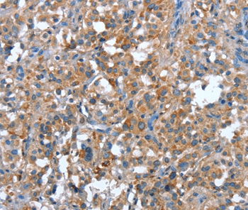
Anti-PLA2G6 Antibody Picoband(r)
A01648-1-CARRIER-FREE
ApplicationsImmunoFluorescence, Western Blot, ELISA, ImmunoCytoChemistry
Product group Antibodies
ReactivityHuman, Mouse, Rat
TargetPLA2G6
Overview
- SupplierBoster Bio
- Product NameAnti-PLA2G6 Antibody Picoband(r)
- Delivery Days Customer9
- ApplicationsImmunoFluorescence, Western Blot, ELISA, ImmunoCytoChemistry
- CertificationResearch Use Only
- ClonalityPolyclonal
- Concentration500 ug/ml
- Gene ID8398
- Target namePLA2G6
- Target descriptionphospholipase A2 group VI
- Target synonymsCaI-PLA2, GVI, INAD1, IPLA2-VIA, NBIA2, NBIA2A, NBIA2B, PARK14, PLA2, PNPLA9, iPLA2, iPLA2beta, 85/88 kDa calcium-independent phospholipase A2, 2-lysophosphatidylcholine acylhydrolase, 85 kDa calcium-independent phospholipase A2, GVI PLA2, iPLA2-beta, intracellular membrane-associated calcium-independent phospholipase A2 beta, neurodegeneration with brain iron accumulation 2, palmitoyl-CoA hydrolase, patatin-like phospholipase domain-containing protein 9, phospholipase A2, group VI (cytosolic, calcium-independent)
- HostRabbit
- IsotypeIgG
- Protein IDO60733
- Protein Name85/88 kDa calcium-independent phospholipase A2
- Scientific DescriptionBoster Bio Anti-PLA2G6 Antibody Picoband® catalog # A01648-1. Tested in ELISA, ICC/IF, WB applications. This antibody reacts with Human, Mouse, Rat. The brand Picoband indicates this is a premium antibody that guarantees superior quality, high affinity, and strong signals with minimal background in Western blot applications. Only our best-performing antibodies are designated as Picoband, ensuring unmatched performance.
- ReactivityHuman, Mouse, Rat
- Storage Instruction-20°C,2°C to 8°C
- UNSPSC12352203
Related products
Product group Antibodies
PLA2G6 AntibodyCSB-PA060291
ApplicationsWestern Blot, ELISA
ReactivityHuman, Mouse, Rat
TargetPLA2G6
- SizePrice
Product group Antibodies
Anti-PLA2G6 Antibody144-66764
ApplicationsWestern Blot
ReactivityHuman, Rat
TargetPLA2G6
- SizePrice
Product group Antibodies
Anti-PLA2G6 AntibodyA45552
ApplicationsImmunoHistoChemistry
ReactivityHuman, Mouse, Rat
- SizePrice
Product group Antibodies

PLA2G6 / IPLA2 AntibodyLS-C749788
ApplicationsWestern Blot
ReactivityHuman, Mouse
TargetPLA2G6
- SizePrice

Product group Antibodies
Anti-PLA2G6 AntibodyHPA001171
ApplicationsWestern Blot, ImmunoCytoChemistry
ReactivityHuman
TargetPLA2G6
- SizePrice
![Drosophila tissue extract (50 μg) was separated by 7.5% SDS-PAGE, and the membrane was blotted with PLA2G6 antibody [N3C3] (GTX103717) diluted at 1:500. The HRP-conjugated anti-rabbit IgG antibody (GTX213110-01) was used to detect the primary antibody.](https://www.genetex.com/upload/website/prouct_img/normal/GTX103717/GTX103717_40625_20230324_WB_Drosophila_brain_23032819_118.webp)
Product group Antibodies
PLA2G6 antibody [N3C3]GTX103717
ApplicationsWestern Blot
ReactivityDrosophila, Human
TargetPLA2G6
- SizePrice
Product group Antibodies
CaIPLA2 Polyclonal AntibodyBS-22878R
ApplicationsImmunoFluorescence, ImmunoCytoChemistry, ImmunoHistoChemistry, ImmunoHistoChemistry Frozen, ImmunoHistoChemistry Paraffin
ReactivityBovine, Canine, Human, Mouse, Porcine, Rabbit, Rat, Sheep
TargetPLA2G6
- SizePrice
Product group Antibodies
ApplicationsWestern Blot, ELISA, ImmunoCytoChemistry, ImmunoHistoChemistry, ImmunoHistoChemistry Frozen, ImmunoHistoChemistry Paraffin
ReactivityMouse, Rat
TargetPLA2G6
- SizePrice