


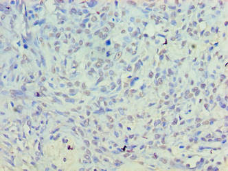
Immunohistochemical staining of human duodenum shows distinct granular cytoplasmic positivity in glandular cells.

Immunohistochemical staining of human duodenum shows distinct granular cytoplasmic positivity in glandular cells.
Anti-PLD5 Antibody
HPA028389
ApplicationsImmunoCytoChemistry, ImmunoHistoChemistry
Product group Antibodies
ReactivityHuman
TargetPLD5
Overview
- SupplierAtlas Antibodies
- Product NameAnti-PLD5 Antibody
- Delivery Days Customer4
- ApplicationsImmunoCytoChemistry, ImmunoHistoChemistry
- CertificationResearch Use Only
- ClonalityPolyclonal
- ConjugateUnconjugated
- Gene ID200150
- Target namePLD5
- Target descriptionphospholipase D family member 5
- Target synonymsPLDC, inactive phospholipase D5, inactive PLD 5, inactive choline phosphatase 5, inactive phosphatidylcholine-hydrolyzing phospholipase D5
- HostRabbit
- IsotypeIgG
- Protein IDQ8N7P1
- Protein NameInactive phospholipase D5
- Scientific DescriptionRecombinant Protein Epitope Signature Tag (PrEST) antigen sequence
- ReactivityHuman
- Storage Instruction-20°C,2°C to 8°C
- UNSPSC41116161
Datasheet
MSDS
Related products
Product group Antibodies
PLD5 AntibodyCSB-PA836686LA01HU
ApplicationsELISA, ImmunoHistoChemistry
ReactivityHuman
TargetPLD5
- SizePrice

Product group Antibodies
Anti-PLD5 Antibody Picoband(r)A14197-1-CARRIER-FREE
ApplicationsFlow Cytometry, Western Blot, ELISA, ImmunoHistoChemistry
ReactivityHuman, Mouse, Rat
TargetPLD5
- SizePrice
Product group Antibodies
PLD5 AntibodyPACO38554
ApplicationsELISA, ImmunoHistoChemistry
ReactivityHuman
TargetPLD5
- SizePrice
Product group Antibodies

PLD5 / Phospholipase D5 Antibody (HRP)LS-C318328
ApplicationsWestern Blot, ELISA
ReactivityHuman
TargetPLD5
- SizePrice

Product group Antibodies
Anti-PLD5 AntibodyHPA050367
ApplicationsImmunoCytoChemistry
ReactivityHuman
TargetPLD5
- SizePrice