


Immunofluorescent staining of human cell line HEL shows localization to nucleoli.
Immunofluorescent staining of human cell line HEL shows localization to nucleoli.
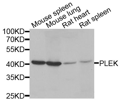
Anti-PLEK Antibody
HPA057341
ApplicationsImmunoCytoChemistry
Product group Antibodies
ReactivityHuman
TargetPLEK
Overview
- SupplierAtlas Antibodies
- Product NameAnti-PLEK Antibody
- Delivery Days Customer4
- ApplicationsImmunoCytoChemistry
- CertificationResearch Use Only
- ClonalityPolyclonal
- ConjugateUnconjugated
- Gene ID5341
- Target namePLEK
- Target descriptionpleckstrin
- Target synonymsP47, PLEK1, pleckstrin, platelet 47 kDa protein
- HostRabbit
- IsotypeIgG
- Protein IDP08567
- Protein NamePleckstrin
- Scientific DescriptionRecombinant Protein Epitope Signature Tag (PrEST) antigen sequence
- ReactivityHuman
- Storage Instruction-20°C,2°C to 8°C
- UNSPSC41116161
Datasheet
MSDS
Related products
Product group Antibodies
Anti-PLEK AntibodyA31282
ApplicationsWestern Blot, ImmunoHistoChemistry
ReactivityHuman, Mouse
- SizePrice

Product group Antibodies
Anti-Pleckstrin/PLEK Antibody Picoband(r)A06656-3-CARRIER-FREE
ApplicationsFlow Cytometry, Western Blot, ELISA, ImmunoHistoChemistry
ReactivityHuman
TargetPLEK
- SizePrice
Product group Antibodies
Anti-PLEK Antibody144-06305
ApplicationsWestern Blot, ImmunoHistoChemistry
ReactivityHuman, Mouse, Rat
TargetPLEK
- SizePrice
Product group Antibodies

Pleckstrin AntibodyABX433144
ApplicationsFlow Cytometry, ImmunoFluorescence, Western Blot, ELISA, ImmunoCytoChemistry, ImmunoHistoChemistry
- SizePrice
Product group Antibodies

PLEK / Pleckstrin AntibodyLS-C748391
ApplicationsWestern Blot, ImmunoHistoChemistry
ReactivityHuman
TargetPLEK
- SizePrice
Product group Antibodies
ApplicationsImmunoFluorescence, Western Blot, ImmunoHistoChemistry, ImmunoHistoChemistry Frozen, ImmunoHistoChemistry Paraffin
ReactivityHuman, Mouse, Rat
TargetPLEK
- SizePrice
Product group Antibodies

Goat anti-PleckstrinEB06169
ApplicationsFlow Cytometry, ImmunoFluorescence, Western Blot, ELISA, ImmunoHistoChemistry
ReactivityHuman
TargetPLEK
- SizePrice
Product group Antibodies
PLEK AntibodyCSB-PA018152LA01HU
ApplicationsELISA
ReactivityHuman
TargetPLEK
- SizePrice