

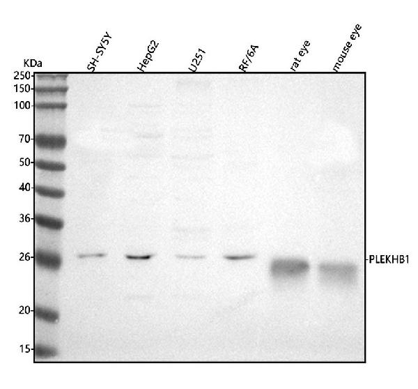
Figure 1. Western blot analysis of PLEKHB1 using anti-PLEKHB1 antibody (A11037-1). Electrophoresis was performed on a 5-20% SDS-PAGE gel at 70V (Stacking gel) / 90V (Resolving gel) for 2-3 hours. The sample well of each lane was loaded with 30 ug of sample under reducing conditions. Lane 1: human SH-SY5Y whole cell lysates, Lane 2: human HepG2 whole cell lysates, Lane 3: human U251 whole cell lysates, Lane 4: monkey RF/6A whole cell lysates, Lane 5: rat eye tissue lysates, Lane 6: mouse eye tissue lysates. After electrophoresis, proteins were transferred to a nitrocellulose membrane at 150 mA for 50-90 minutes. Blocked the membrane with 5% non-fat milk/TBS for 1.5 hour at RT. The membrane was incubated with rabbit anti-PLEKHB1 antigen affinity purified polyclonal antibody (Catalog # A11037-1) at 0.5 microg/mL overnight at 4°C, then washed with TBS-0.1%Tween 3 times with 5 minutes each and probed with a goat anti-rabbit IgG-HRP secondary antibody at a dilution of 1:5000 for 1.5 hour at RT. The signal is developed using an Enhanced Chemiluminescent detection (ECL) kit (Catalog # EK1002) with Tanon 5200 system. A specific band was detected for PLEKHB1 at approximately 27 kDa. The expected band size for PLEKHB1 is at 27 kDa.
Figure 1. Western blot analysis of PLEKHB1 using anti-PLEKHB1 antibody (A11037-1). Electrophoresis was performed on a 5-20% SDS-PAGE gel at 70V (Stacking gel) / 90V (Resolving gel) for 2-3 hours. The sample well of each lane was loaded with 30 ug of sample under reducing conditions. Lane 1: human SH-SY5Y whole cell lysates, Lane 2: human HepG2 whole cell lysates, Lane 3: human U251 whole cell lysates, Lane 4: monkey RF/6A whole cell lysates, Lane 5: rat eye tissue lysates, Lane 6: mouse eye tissue lysates. After electrophoresis, proteins were transferred to a nitrocellulose membrane at 150 mA for 50-90 minutes. Blocked the membrane with 5% non-fat milk/TBS for 1.5 hour at RT. The membrane was incubated with rabbit anti-PLEKHB1 antigen affinity purified polyclonal antibody (Catalog # A11037-1) at 0.5 microg/mL overnight at 4°C, then washed with TBS-0.1%Tween 3 times with 5 minutes each and probed with a goat anti-rabbit IgG-HRP secondary antibody at a dilution of 1:5000 for 1.5 hour at RT. The signal is developed using an Enhanced Chemiluminescent detection (ECL) kit (Catalog # EK1002) with Tanon 5200 system. A specific band was detected for PLEKHB1 at approximately 27 kDa. The expected band size for PLEKHB1 is at 27 kDa.
Anti-PLEKHB1 Antibody Picoband(r)
A11037-1-CARRIER-FREE
ApplicationsWestern Blot, ELISA
Product group Antibodies
ReactivityHuman, Monkey, Mouse, Rat
TargetPLEKHB1
Overview
- SupplierBoster Bio
- Product NameAnti-PLEKHB1 Antibody Picoband(r)
- Delivery Days Customer9
- ApplicationsWestern Blot, ELISA
- CertificationResearch Use Only
- ClonalityPolyclonal
- Concentration500 ug/ml
- Gene ID58473
- Target namePLEKHB1
- Target descriptionpleckstrin homology domain containing B1
- Target synonymsKPL1, PHR1, PHRET1, pleckstrin homology domain-containing family B member 1, PH domain containing, retinal 1, PH domain-containing family B member 1, PH domain-containing protein in retina 1, evectin-1, pleckstrin homology domain containing, family B (evectins) member 1, pleckstrin homology domain retinal protein 1
- HostRabbit
- IsotypeIgG
- Protein IDQ9UF11
- Protein NamePleckstrin homology domain-containing family B member 1
- Scientific DescriptionBoster Bio Anti-PLEKHB1 Antibody Picoband® catalog # A11037-1. Tested in WB, ELISA applications. This antibody reacts with Human, Monkey, Mouse, Rat. The brand Picoband indicates this is a premium antibody that guarantees superior quality, high affinity, and strong signals with minimal background in Western blot applications. Only our best-performing antibodies are designated as Picoband, ensuring unmatched performance.
- ReactivityHuman, Monkey, Mouse, Rat
- Storage Instruction-20°C,2°C to 8°C
- UNSPSC12352203
Related products

Product group Antibodies
PHR1 AntibodyCSB-PA165910XA01OFG
ApplicationsWestern Blot, ELISA
ReactivityPlant
- SizePrice
Product group Antibodies
PHR1 antibody, N-termGTX88536
ApplicationsWestern Blot
ReactivityHuman, Rat
TargetPLEKHB1
- SizePrice
Product group Antibodies

PLEKHB1 AntibodyLS-C752077
ApplicationsWestern Blot, ELISA
ReactivityHuman
TargetPLEKHB1
- SizePrice