


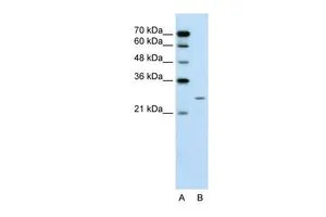
Figure 1. Western blot analysis of Plunc/BPIFA1 using anti-Plunc/BPIFA1 antibody (A03162-2). Electrophoresis was performed on a 5-20% SDS-PAGE gel at 70V (Stacking gel) / 90V (Resolving gel) for 2-3 hours. The sample well of each lane was loaded with 30 ug of sample under reducing conditions. Lane 1: mouse NIH/3T3 whole cell lysates. After electrophoresis, proteins were transferred to a nitrocellulose membrane at 150 mA for 50-90 minutes. Blocked the membrane with 5% non-fat milk/TBS for 1.5 hour at RT. The membrane was incubated with rabbit anti-Plunc/BPIFA1 antigen affinity purified polyclonal antibody (Catalog # A03162-2) at 0.5 microg/mL overnight at 4°C, then washed with TBS-0.1%Tween 3 times with 5 minutes each and probed with a goat anti-rabbit IgG-HRP secondary antibody at a dilution of 1:5000 for 1.5 hour at RT. The signal is developed using an Enhanced Chemiluminescent detection (ECL) kit (Catalog # EK1002) with Tanon 5200 system. A specific band was detected for Plunc/BPIFA1 at approximately 27 kDa. The expected band size for Plunc/BPIFA1 is at 27 kDa.
Figure 1. Western blot analysis of Plunc/BPIFA1 using anti-Plunc/BPIFA1 antibody (A03162-2). Electrophoresis was performed on a 5-20% SDS-PAGE gel at 70V (Stacking gel) / 90V (Resolving gel) for 2-3 hours. The sample well of each lane was loaded with 30 ug of sample under reducing conditions. Lane 1: mouse NIH/3T3 whole cell lysates. After electrophoresis, proteins were transferred to a nitrocellulose membrane at 150 mA for 50-90 minutes. Blocked the membrane with 5% non-fat milk/TBS for 1.5 hour at RT. The membrane was incubated with rabbit anti-Plunc/BPIFA1 antigen affinity purified polyclonal antibody (Catalog # A03162-2) at 0.5 microg/mL overnight at 4°C, then washed with TBS-0.1%Tween 3 times with 5 minutes each and probed with a goat anti-rabbit IgG-HRP secondary antibody at a dilution of 1:5000 for 1.5 hour at RT. The signal is developed using an Enhanced Chemiluminescent detection (ECL) kit (Catalog # EK1002) with Tanon 5200 system. A specific band was detected for Plunc/BPIFA1 at approximately 27 kDa. The expected band size for Plunc/BPIFA1 is at 27 kDa.
Anti-Plunc/BPIFA1 Antibody Picoband(r)
A03162-2-CARRIER-FREE
ApplicationsWestern Blot, ELISA
Product group Antibodies
ReactivityHuman, Mouse
TargetBPIFA1
Overview
- SupplierBoster Bio
- Product NameAnti-Plunc/BPIFA1 Antibody Picoband(r)
- Delivery Days Customer9
- ApplicationsWestern Blot, ELISA
- CertificationResearch Use Only
- ClonalityPolyclonal
- Concentration500 ug/ml
- Gene ID51297
- Target nameBPIFA1
- Target descriptionBPI fold containing family A member 1
- Target synonymsLUNX, NASG, PLUNC, SPLUNC1, SPURT, bA49G10.5, BPI fold-containing family A member 1, ligand-binding protein RYA3, lung-specific protein X, nasopharyngeal carcinoma-related protein, palate lung and nasal epithelium clone protein, palate, lung and nasal epithelium associated, protein Plunc, secretory protein in upper respiratory tracts, short PLUNC1, tracheal epithelium enriched protein, von Ebner protein Hl
- HostRabbit
- IsotypeIgG
- Protein IDQ9NP55
- Protein NameBPI fold-containing family A member 1
- Scientific DescriptionBoster Bio Anti-Plunc/BPIFA1 Antibody Picoband® catalog # A03162-2. Tested in ELISA, WB applications. This antibody reacts with Human, Mouse. The brand Picoband indicates this is a premium antibody that guarantees superior quality, high affinity, and strong signals with minimal background in Western blot applications. Only our best-performing antibodies are designated as Picoband, ensuring unmatched performance.
- ReactivityHuman, Mouse
- Storage Instruction-20°C,2°C to 8°C
- UNSPSC12352203
Related products
Product group Antibodies
Anti-BPIFA1 AntibodyA28742
ApplicationsWestern Blot
ReactivityHuman, Rat
- SizePrice
Product group Antibodies
Anti-BPIFA1 Antibody144-08634
ApplicationsWestern Blot
ReactivityHuman, Mouse, Rat
TargetBPIFA1
- SizePrice
Product group Antibodies

Goat anti-BPIFA1 / PLUNCEB11481
ApplicationsWestern Blot, ELISA
ReactivityHuman
TargetBPIFA1
- SizePrice
Product group Antibodies
ApplicationsImmunoPrecipitation, Western Blot, ImmunoCytoChemistry, ImmunoHistoChemistry
TargetBPIFA1
- SizePrice
Product group Antibodies
BPIFA1 AntibodyCSB-PA020281
ApplicationsELISA, ImmunoHistoChemistry
ReactivityHuman
TargetBPIFA1
- SizePrice
Product group Antibodies

BPIFA1 / SPLUNC1 AntibodyLS-C410168
ApplicationsWestern Blot
ReactivityHuman, Mouse, Rat
TargetBPIFA1
- SizePrice
Product group Antibodies
PLUNC antibody, InternalGTX47015
ApplicationsWestern Blot, ImmunoHistoChemistry, ImmunoHistoChemistry Paraffin
ReactivityHuman
TargetBPIFA1
- SizePrice
Product group Antibodies
Anti-BPIFA1 AntibodyCAB8634
ApplicationsWestern Blot, ELISA
ReactivityMouse
TargetBPIFA1
- SizePrice