


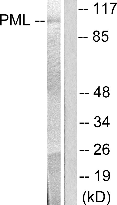
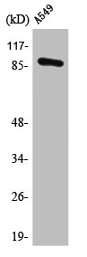
Figure 1. Western blot analysis of PML using anti-PML antibody (PB10084). Electrophoresis was performed on a 5-20% SDS-PAGE gel at 70V (Stacking gel) / 90V (Resolving gel) for 2-3 hours. The sample well of each lane was loaded with 30 ug of sample under reducing conditions. Lane 1: human Hela whole cell lysates, Lane 2: human HEK293 whole cell lysates, Lane 3: human U87 whole cell lysates, Lane 4: human A431 whole cell lysates, Lane 5: human K562 whole cell lysates. red to a nitrocellulose membrane at 150 mA for 50-90 minutes. Blocked the membrane with 5% non-fat milk/TBS for 1.5 hour at RT. The membrane was incubated with rabbit anti-PML antigen affinity purified polyclonal antibody (Catalog # PB10084) at 0.5 microg/mL overnight at 4°C, then washed with TBS-0.1%Tween 3 times with 5 minutes each and probed with a goat anti-rabbit IgG-HRP secondary antibody at a dilution of 1:5000 for 1.5 hour at RT. The signal is developed using an Enhanced Chemiluminescent detection (ECL) kit (Catalog # EK1002) with Tanon 5200 system. A specific band was detected for PML at approximately 90-160 kDa. The expected band size for PML is at 98 kDa.
. PML was detected in paraffin-embedded section of human intestinal cancer tissue. Heat mediated antigen retrieval was performed in EDTA buffer (pH8.0, epitope retrieval solution). The tissue section was blocked with 10% goat serum. The tissue section was then incubated with 1microg/ml rabbit anti-PML Antibody (PB10084) overnight at 4°C. Biotinylated goat anti-rabbit IgG was used as secondary antibody and incubated for 30 minutes at 37°C. The tissue section was developed using Strepavidin-Biotin-Complex (SABC) (Catalog # SA1022) with DAB as the chromogen.")
. Overlay histogram showing A431 cells stained with PB10084 (Blue line). To facilitate intracellular staining, cells were fixed with 4% paraformaldehyde and permeabilized with permeabilization buffer. The cells were blocked with 10% normal goat serum. And then incubated with rabbit anti-PML Antibody (PB10084,1microg/1x106 cells) for 30 min at 20°C. DyLight®488 conjugated goat anti-rabbit IgG (BA1127, 5-10microg/1x106 cells) was used as secondary antibody for 30 minutes at 20°C. Isotype control antibody (Green line) was rabbit IgG (1microg/1x106) used under the same conditions. Unlabelled sample without incubation with primary antibody and secondary antibody (Red line) was used as a blank control.")
. PML was detected in paraffin-embedded section of human mammary cancer tissue. Heat mediated antigen retrieval was performed in EDTA buffer (pH8.0, epitope retrieval solution). The tissue section was blocked with 10% goat serum. The tissue section was then incubated with 1microg/ml rabbit anti-PML Antibody (PB10084) overnight at 4°C. Biotinylated goat anti-rabbit IgG was used as secondary antibody and incubated for 30 minutes at 37°C. The tissue section was developed using Strepavidin-Biotin-Complex (SABC) (Catalog # SA1022) with DAB as the chromogen.")
Figure 1. Western blot analysis of PML using anti-PML antibody (PB10084). Electrophoresis was performed on a 5-20% SDS-PAGE gel at 70V (Stacking gel) / 90V (Resolving gel) for 2-3 hours. The sample well of each lane was loaded with 30 ug of sample under reducing conditions. Lane 1: human Hela whole cell lysates, Lane 2: human HEK293 whole cell lysates, Lane 3: human U87 whole cell lysates, Lane 4: human A431 whole cell lysates, Lane 5: human K562 whole cell lysates. red to a nitrocellulose membrane at 150 mA for 50-90 minutes. Blocked the membrane with 5% non-fat milk/TBS for 1.5 hour at RT. The membrane was incubated with rabbit anti-PML antigen affinity purified polyclonal antibody (Catalog # PB10084) at 0.5 microg/mL overnight at 4°C, then washed with TBS-0.1%Tween 3 times with 5 minutes each and probed with a goat anti-rabbit IgG-HRP secondary antibody at a dilution of 1:5000 for 1.5 hour at RT. The signal is developed using an Enhanced Chemiluminescent detection (ECL) kit (Catalog # EK1002) with Tanon 5200 system. A specific band was detected for PML at approximately 90-160 kDa. The expected band size for PML is at 98 kDa.
Anti-PML Protein Antibody Picoband(r)
PB10084-DYLIGHT594
ApplicationsFlow Cytometry, Western Blot, ImmunoHistoChemistry
Product group Antibodies
ReactivityHuman
TargetPML
Overview
- SupplierBoster Bio
- Product NameAnti-PML Protein Antibody Picoband(r)
- Delivery Days Customer9
- Application Supplier NoteTested Species: In-house tested species with positive results. By Heat: Boiling the paraffin sections in 10mM citrate buffer, pH6.0, for 20mins is required for the staining of formalin/paraffin sections. Other applications have not been tested. Optimal dilutions should be determined by end users.
- ApplicationsFlow Cytometry, Western Blot, ImmunoHistoChemistry
- CertificationResearch Use Only
- ClonalityPolyclonal
- Concentration500 ug/ml
- ConjugateOther Conjugate
- Gene ID5371
- Target namePML
- Target descriptionPML nuclear body scaffold
- Target synonymsMYL, PP8675, RNF71, TRIM19, protein PML, E3 SUMO-protein ligase PML, PML/RARA fusion, RING finger protein 71, RING-type E3 SUMO transferase PML, probable transcription factor PML, promyelocytic leukemia protein, promyelocytic leukemia, inducer of, tripartite motif protein TRIM19, tripartite motif-containing protein 19
- HostRabbit
- IsotypeIgG
- Protein IDP29590
- Protein NameProtein PML
- Scientific DescriptionBoster Bio Anti-PML Protein Antibody Picoband® catalog # PB10084. Tested in Flow Cytometry, IHC, WB applications. This antibody reacts with Human. The brand Picoband indicates this is a premium antibody that guarantees superior quality, high affinity, and strong signals with minimal background in Western blot applications. Only our best-performing antibodies are designated as Picoband, ensuring unmatched performance.
- ReactivityHuman
- Storage Instruction-20°C,2°C to 8°C
- UNSPSC12352203
Related products
Product group Antibodies
Anti-PML Antibody144-65308
ApplicationsWestern Blot
ReactivityHuman, Mouse, Rat
TargetPML
- SizePrice
Product group Antibodies
ApplicationsFlow Cytometry
TargetPML
- SizePrice
Product group Antibodies
Anti-PML AntibodyA99234
ApplicationsImmunoFluorescence, Western Blot, ELISA, ImmunoHistoChemistry
ReactivityHuman
- SizePrice
Product group Antibodies
Anti-PML Protein Antibody Picoband(r)PB10084-CARRIER-FREE
ApplicationsFlow Cytometry, Western Blot, ImmunoHistoChemistry
ReactivityHuman
TargetPML
- SizePrice
Product group Antibodies
PML AntibodyCSB-PA003823
ApplicationsImmunoFluorescence, Western Blot, ELISA, ImmunoHistoChemistry
ReactivityHuman
TargetPML
- SizePrice
Product group Antibodies
PML antibody [N1N2], N-termGTX112996
ApplicationsImmunoPrecipitation, Western Blot
ReactivityHuman
TargetPML
- SizePrice
Product group Antibodies
PML Recombinant Antibody, AbBy Fluor-594 ConjugatedBSM-61469R-BF594
ApplicationsFlow Cytometry, ImmunoFluorescence, Western Blot, ImmunoCytoChemistry
ReactivityHuman, Mouse, Rat
TargetPML
- SizePrice

Product group Antibodies
Anti-PML AntibodyHPA008312
ApplicationsChIP Chromatin ImmunoPrecipitation, ImmunoHistoChemistry
ReactivityHuman
TargetPML
- SizePrice