


Immunofluorescent staining of human cell line CACO-2 shows localization to nucleoplasm.
 shows similar pattern to independent antibody HPA068992 (B).")
Immunofluorescent staining of human cell line CACO-2 shows localization to nucleoplasm.
Anti-POLR2E Antibody
HPA063030
ApplicationsWestern Blot, ImmunoCytoChemistry
Product group Antibodies
ReactivityHuman
TargetPOLR2E
Overview
- SupplierAtlas Antibodies
- Product NameAnti-POLR2E Antibody
- Delivery Days Customer4
- ApplicationsWestern Blot, ImmunoCytoChemistry
- CertificationResearch Use Only
- ClonalityPolyclonal
- ConjugateUnconjugated
- Gene ID5434
- Target namePOLR2E
- Target descriptionRNA polymerase II, I and III subunit E
- Target synonymsRPABC1, RPB5, XAP4, hRPB25, hsRPB5, DNA-directed RNA polymerases I, II, and III subunit RPABC1, DNA directed RNA polymerase II 23 kda polypeptide, DNA-directed RNA polymerase II 23 kDa polypeptide, DNA-directed RNA polymerase II subunit E, DNA-directed RNA polymerase subunit RPABC1, RNA polymerase II subunit E, RNA polymerases I, II, and III subunit ABC1, RPB5 homolog, polymerase (RNA) II (DNA directed) polypeptide E, 25kDa, polymerase (RNA) II subunit E
- HostRabbit
- IsotypeIgG
- Protein IDP19388
- Protein NameDNA-directed RNA polymerases I, II, and III subunit RPABC1
- Scientific DescriptionRecombinant Protein Epitope Signature Tag (PrEST) antigen sequence
- ReactivityHuman
- Storage Instruction-20°C,2°C to 8°C
- UNSPSC41116161
Datasheet
MSDS
Related products
Product group Antibodies
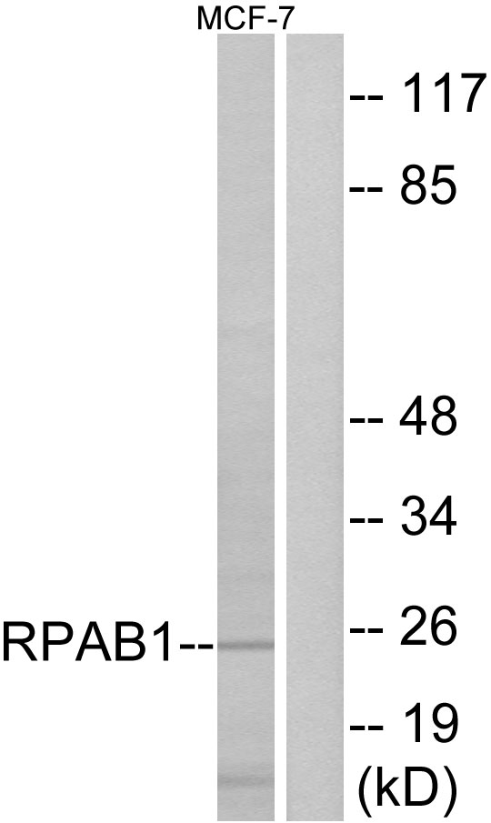
Anti-RPAB1 AntibodyA97283
ApplicationsWestern Blot, ELISA
ReactivityHuman, Mouse, Rat
- SizePrice

Product group Antibodies
Anti-RPB5/POLR2E Antibody Picoband(r)A08765-1-CARRIER-FREE
ApplicationsFlow Cytometry, ImmunoFluorescence, Western Blot, ELISA, ImmunoCytoChemistry
ReactivityHuman, Rat
TargetPOLR2E
- SizePrice
Product group Antibodies
Anti-POLR2E Antibody144-01755
ApplicationsImmunoFluorescence, Western Blot, ImmunoHistoChemistry
ReactivityHuman, Mouse
TargetPOLR2E
- SizePrice

Product group Antibodies
POLR2E AntibodyCSB-PA018332LA01HU
ApplicationsELISA
ReactivityHuman
TargetPOLR2E
- SizePrice
Product group Antibodies

POLR2E AntibodyLS-C331669
ApplicationsImmunoFluorescence, Western Blot, ImmunoHistoChemistry
ReactivityHuman, Mouse
TargetPOLR2E
- SizePrice

Product group Antibodies
Anti-POLR2E AntibodyHPA068992
ApplicationsWestern Blot, ImmunoHistoChemistry
ReactivityHuman
TargetPOLR2E
- SizePrice