


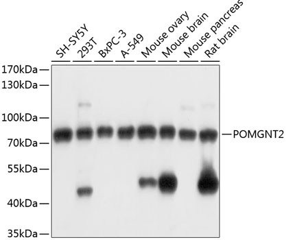
Figure 1. Western blot analysis of POMGNT2 using anti-POMGNT2 antibody (A11987-1). Electrophoresis was performed on a 5-20% SDS-PAGE gel at 70V (Stacking gel) / 90V (Resolving gel) for 2-3 hours. The sample well of each lane was loaded with 30 ug of sample under reducing conditions. Lane 1: human 293T whole cell lysates, Lane 2: human PC-3 whole cell lysates, Lane 3: human Jurkat whole cell lysates, Lane 4: human U2OS whole cell lysates, Lane 5: rat brain tissue lysates, Lane 6: mouse brain tissue lysates, Lane 7: mouse pancreas tissue lysates. After electrophoresis, proteins were transferred to a nitrocellulose membrane at 150 mA for 50-90 minutes. Blocked the membrane with 5% non-fat milk/TBS for 1.5 hour at RT. The membrane was incubated with rabbit anti-POMGNT2 antigen affinity purified polyclonal antibody (Catalog # A11987-1) at 0.5 microg/mL overnight at 4°C, then washed with TBS-0.1%Tween 3 times with 5 minutes each and probed with a goat anti-rabbit IgG-HRP secondary antibody at a dilution of 1:5000 for 1.5 hour at RT. The signal is developed using an Enhanced Chemiluminescent detection (ECL) kit (Catalog # EK1002) with Tanon 5200 system. A specific band was detected for POMGNT2 at approximately 75 kDa. The expected band size for POMGNT2 is at 67 kDa.
Figure 1. Western blot analysis of POMGNT2 using anti-POMGNT2 antibody (A11987-1). Electrophoresis was performed on a 5-20% SDS-PAGE gel at 70V (Stacking gel) / 90V (Resolving gel) for 2-3 hours. The sample well of each lane was loaded with 30 ug of sample under reducing conditions. Lane 1: human 293T whole cell lysates, Lane 2: human PC-3 whole cell lysates, Lane 3: human Jurkat whole cell lysates, Lane 4: human U2OS whole cell lysates, Lane 5: rat brain tissue lysates, Lane 6: mouse brain tissue lysates, Lane 7: mouse pancreas tissue lysates. After electrophoresis, proteins were transferred to a nitrocellulose membrane at 150 mA for 50-90 minutes. Blocked the membrane with 5% non-fat milk/TBS for 1.5 hour at RT. The membrane was incubated with rabbit anti-POMGNT2 antigen affinity purified polyclonal antibody (Catalog # A11987-1) at 0.5 microg/mL overnight at 4°C, then washed with TBS-0.1%Tween 3 times with 5 minutes each and probed with a goat anti-rabbit IgG-HRP secondary antibody at a dilution of 1:5000 for 1.5 hour at RT. The signal is developed using an Enhanced Chemiluminescent detection (ECL) kit (Catalog # EK1002) with Tanon 5200 system. A specific band was detected for POMGNT2 at approximately 75 kDa. The expected band size for POMGNT2 is at 67 kDa.
Anti-POMGNT2 Antibody Picoband(r)
A11987-1-CY3
ApplicationsWestern Blot, ELISA
Product group Antibodies
ReactivityHuman, Mouse, Rat
TargetPOMGNT2
Overview
- SupplierBoster Bio
- Product NameAnti-POMGNT2 Antibody Picoband(r)
- Delivery Days Customer9
- ApplicationsWestern Blot, ELISA
- CertificationResearch Use Only
- ClonalityPolyclonal
- Concentration500 ug/ml
- ConjugateCy3
- Gene ID84892
- Target namePOMGNT2
- Target descriptionprotein O-linked mannose N-acetylglucosaminyltransferase 2 (beta 1,4-)
- Target synonymsAGO61, C3orf39, GTDC2, MDDGA8, MDDGC8, protein O-linked-mannose beta-1,4-N-acetylglucosaminyltransferase 2, glycosyltransferase-like domain containing 2, glycosyltransferase-like domain-containing protein 2, protein O-mannose beta-1,4-N-acetylglucosaminyltransferase 2
- HostRabbit
- Protein IDQ8NAT1
- Protein NameProtein O-linked-mannose beta-1,4-N-acetylglucosaminyltransferase 2
- Scientific DescriptionBoster Bio Anti-POMGNT2 Antibody Picoband® catalog # A11987-1. Tested in WB, ELISA applications. This antibody reacts with Human, Mouse, Rat. The brand Picoband indicates this is a premium antibody that guarantees superior quality, high affinity, and strong signals with minimal background in Western blot applications. Only our best-performing antibodies are designated as Picoband, ensuring unmatched performance.
- ReactivityHuman, Mouse, Rat
- Storage Instruction-20°C,2°C to 8°C
- UNSPSC12352203
Related products
Product group Antibodies
Anti-POMGNT2 AntibodyA91128
ApplicationsWestern Blot
ReactivityHuman, Mouse, Rat
- SizePrice
Product group Antibodies
C3orf39 Polyclonal AntibodyBS-9830R
ApplicationsImmunoFluorescence, Western Blot, ImmunoHistoChemistry, ImmunoHistoChemistry Paraffin
ReactivityHuman, Mouse, Rat
TargetPOMGNT2
- SizePrice
Product group Antibodies
Anti-POMGNT2 Antibody144-60691
ApplicationsWestern Blot
ReactivityHuman, Mouse, Rat
TargetPOMGNT2
- SizePrice
Product group Antibodies

POMGNT2 / GTDC2 AntibodyLS-C749518
ApplicationsWestern Blot
ReactivityHuman, Mouse, Rat
TargetPOMGNT2
- SizePrice
Product group Antibodies
C3ORF39 antibody, InternalGTX46151
ApplicationsWestern Blot
ReactivityHuman
TargetPOMGNT2
- SizePrice

Product group Antibodies
Anti-POMGNT2 AntibodyHPA040989
ApplicationsImmunoHistoChemistry
ReactivityHuman
TargetPOMGNT2
- SizePrice
Product group Antibodies
POMGNT2 AntibodyCSB-PA847672XA01HU
ApplicationsWestern Blot, ELISA
ReactivityHuman
TargetPOMGNT2
- SizePrice
Product group Antibodies
Anti-POMGNT2 Antibody Picoband(r)A11987-1-CARRIER-FREE
ApplicationsWestern Blot, ELISA
ReactivityHuman, Mouse, Rat
TargetPOMGNT2
- SizePrice