


Figure 1. Western blot analysis of POMT2 using anti-POMT2 antibody (A04876-1). Electrophoresis was performed on a 5-20% SDS-PAGE gel at 70V (Stacking gel) / 90V (Resolving gel) for 2-3 hours. The sample well of each lane was loaded with 50ug of sample under reducing conditions. Lane 1: human PC-3 whole cell lysates, Lane 2: human Caco-2 whole cell lysates, Lane 3: human HepG2 whole cell lysates, Lane 4: rat lung tissue lysates, Lane 5: rat testis tissue lysates, Lane 6: rat spleen tissue lysates, Lane 7: mouse lung tissue lysates, Lane 8: mouse testis tissue lysates. After Electrophoresis, proteins were transferred to a Nitrocellulose membrane at 150mA for 50-90 minutes. Blocked the membrane with 5% Non-fat Milk/ TBS for 1.5 hour at RT. The membrane was incubated with rabbit anti-POMT2 antigen affinity purified polyclonal antibody (Catalog # A04876-1) at 0.5 microg/mL overnight at 4°C, then washed with TBS-0.1%Tween 3 times with 5 minutes each and probed with a goat anti-rabbit IgG-HRP secondary antibody at a dilution of 1:10000 for 1.5 hour at RT. The signal is developed using an Enhanced Chemiluminescent detection (ECL) kit (Catalog # EK1002) with Tanon 5200 system. A specific band was detected for POMT2 at approximately 84KD. The expected band size for POMT2 is at 84KD.
Figure 1. Western blot analysis of POMT2 using anti-POMT2 antibody (A04876-1). Electrophoresis was performed on a 5-20% SDS-PAGE gel at 70V (Stacking gel) / 90V (Resolving gel) for 2-3 hours. The sample well of each lane was loaded with 50ug of sample under reducing conditions. Lane 1: human PC-3 whole cell lysates, Lane 2: human Caco-2 whole cell lysates, Lane 3: human HepG2 whole cell lysates, Lane 4: rat lung tissue lysates, Lane 5: rat testis tissue lysates, Lane 6: rat spleen tissue lysates, Lane 7: mouse lung tissue lysates, Lane 8: mouse testis tissue lysates. After Electrophoresis, proteins were transferred to a Nitrocellulose membrane at 150mA for 50-90 minutes. Blocked the membrane with 5% Non-fat Milk/ TBS for 1.5 hour at RT. The membrane was incubated with rabbit anti-POMT2 antigen affinity purified polyclonal antibody (Catalog # A04876-1) at 0.5 microg/mL overnight at 4°C, then washed with TBS-0.1%Tween 3 times with 5 minutes each and probed with a goat anti-rabbit IgG-HRP secondary antibody at a dilution of 1:10000 for 1.5 hour at RT. The signal is developed using an Enhanced Chemiluminescent detection (ECL) kit (Catalog # EK1002) with Tanon 5200 system. A specific band was detected for POMT2 at approximately 84KD. The expected band size for POMT2 is at 84KD.
Anti-POMT2 Antibody Picoband(r)
A04876-1-CARRIER-FREE
ApplicationsFlow Cytometry, ImmunoFluorescence, Western Blot, ImmunoCytoChemistry, ImmunoHistoChemistry
Product group Antibodies
ReactivityHuman, Mouse, Rat
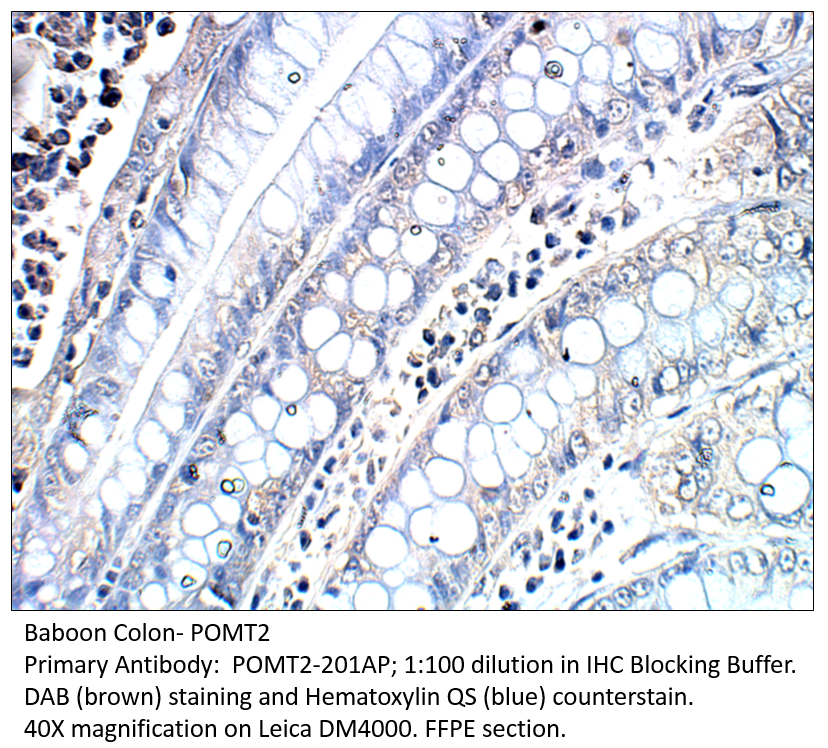
TargetPOMT2
Overview
- SupplierBoster Bio
- Product NameAnti-POMT2 Antibody Picoband(r)
- Delivery Days Customer9
- ApplicationsFlow Cytometry, ImmunoFluorescence, Western Blot, ImmunoCytoChemistry, ImmunoHistoChemistry
- CertificationResearch Use Only
- ClonalityPolyclonal
- Concentration500 ug/ml
- Gene ID29954
- Target namePOMT2
- Target descriptionprotein O-mannosyltransferase 2
- Target synonymsLGMD2N, LGMDR14, MDDGA2, MDDGB2, MDDGC2, protein O-mannosyl-transferase 2, dolichyl-phosphate-mannose--protein mannosyltransferase 2
- HostRabbit
- IsotypeIgG
- Protein IDQ9UKY4
- Protein NameProtein O-mannosyl-transferase 2
- Scientific DescriptionBoster Bio Anti-POMT2 Antibody Picoband® catalog # A04876-1. Tested in Flow Cytometry, IF, IHC, ICC, WB applications. This antibody reacts with Human, Mouse, Rat. The brand Picoband indicates this is a premium antibody that guarantees superior quality, high affinity, and strong signals with minimal background in Western blot applications. Only our best-performing antibodies are designated as Picoband, ensuring unmatched performance.
- ReactivityHuman, Mouse, Rat
- Storage Instruction-20°C,2°C to 8°C
- UNSPSC12352203
Related products
Product group Antibodies
POMT2 AntibodyCSB-PA018367GA01HU
ApplicationsELISA, ImmunoHistoChemistry
ReactivityHuman, Mouse, Rat
TargetPOMT2
- SizePrice
Product group Antibodies
Anti-POMT2 AntibodyA54735
ApplicationsImmunoPrecipitation, Western Blot, ELISA, ImmunoHistoChemistry
ReactivityHuman, Monkey, Mouse
- SizePrice

Product group Antibodies
Anti-POMT2 AntibodyHPA003663
ApplicationsWestern Blot, ImmunoHistoChemistry
ReactivityHuman, Mouse, Rat
TargetPOMT2
- SizePrice
Product group Antibodies

POMT2 Antibody (aa540-567)LS-C158699
ApplicationsWestern Blot
ReactivityHuman
TargetPOMT2
- SizePrice
Product group Antibodies
Anti-POMT2 (C-term) Antibody102-22712
ApplicationsWestern Blot
TargetPOMT2
- SizePrice