


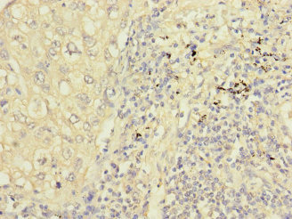
Immunohistochemical staining of human tonsil shows strong nuclear positivity in germinal center cells.

Immunohistochemical staining of human tonsil shows strong nuclear positivity in germinal center cells.
Anti-PPIL2 Antibody
HPA035344
ApplicationsWestern Blot, ImmunoCytoChemistry, ImmunoHistoChemistry
Product group Antibodies
ReactivityHuman
TargetPPIL2
Overview
- SupplierAtlas Antibodies
- Product NameAnti-PPIL2 Antibody
- Delivery Days Customer4
- ApplicationsWestern Blot, ImmunoCytoChemistry, ImmunoHistoChemistry
- CertificationResearch Use Only
- ClonalityPolyclonal
- ConjugateUnconjugated
- Gene ID23759
- Target namePPIL2
- Target descriptionpeptidylprolyl isomerase like 2
- Target synonymsCYC4, CYP60, Cyp-60, UBOX7, hCyP-60, RING-type E3 ubiquitin-protein ligase PPIL2, PPIase, RING-type E3 ubiquitin transferase isomerase-like 2, U-box domain containing 7, cyclophilin, 60kDa, cyclophilin-60, cyclophilin-like protein CyP-60, peptidyl-prolyl cis-trans isomerase-like 2, peptidylprolyl cis-trans isomerase, peptidylprolyl isomerase (cyclophilin)-like 2, probable inactive peptidyl-prolyl cis-trans isomerase-like 2, rotamase PPIL2
- HostRabbit
- IsotypeIgG
- Protein IDQ13356
- Protein NameRING-type E3 ubiquitin-protein ligase PPIL2
- Scientific DescriptionRecombinant Protein Epitope Signature Tag (PrEST) antigen sequence
- ReactivityHuman
- Storage Instruction-20°C,2°C to 8°C
- UNSPSC41116161
Datasheet
MSDS
Related products
Product group Antibodies
Ppil2 Polyclonal AntibodyCAC10912
ApplicationsELISA, ImmunoHistoChemistry
TargetPPIL2
- SizePrice

Product group Antibodies
Anti-PPIL2 AntibodyA11771
ApplicationsWestern Blot
ReactivityHuman, Mouse
- SizePrice
Product group Antibodies
PPIL2 antibodyGTX111086
ApplicationsWestern Blot
ReactivityHuman
TargetPPIL2
- SizePrice
Product group Antibodies
PPIL2 AntibodyCSB-PA621661ESR1HU
ApplicationsELISA, ImmunoHistoChemistry
ReactivityHuman
TargetPPIL2
- SizePrice
Product group Antibodies

PPIL2 / CYP60 AntibodyLS-C676295
ApplicationsELISA, ImmunoHistoChemistry, ImmunoHistoChemistry Paraffin
ReactivityHuman
TargetPPIL2
- SizePrice

Product group Antibodies
Anti-PPIL2 AntibodyHPA055637
ApplicationsImmunoCytoChemistry
ReactivityHuman
TargetPPIL2
- SizePrice