


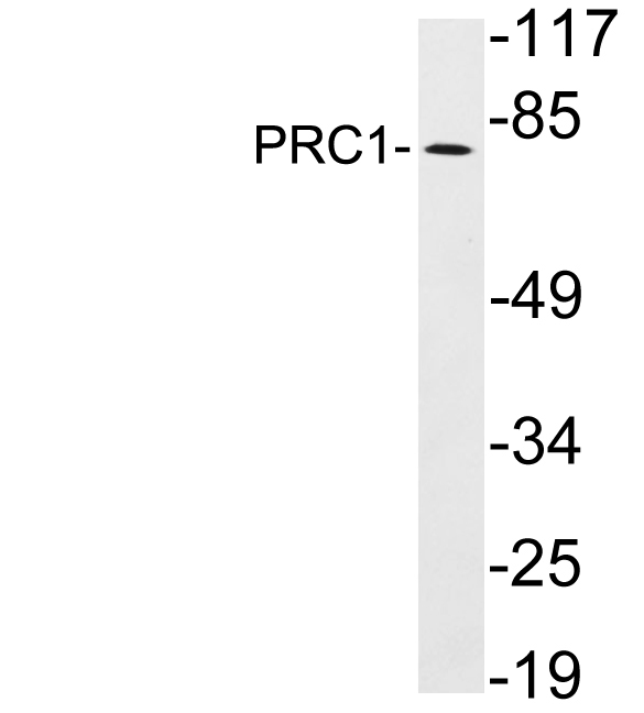
Figure 1. Western blot analysis of PRC1 using anti-PRC1 antibody (PB9814). Electrophoresis was performed on a 5-20% SDS-PAGE gel at 70V (Stacking gel) / 90V (Resolving gel) for 2-3 hours. The sample well of each lane was loaded with 40 ug of sample under reducing conditions. Lane 1: MCF-7 Whole Cell Lysate, Lane 2: SGC Whole Cell Lysate, Lane 3: HELA Whole Cell Lysate. After electrophoresis, proteins were transferred to a nitrocellulose membrane at 150 mA for 50-90 minutes. Blocked the membrane with 5% non-fat milk/TBS for 1.5 hour at RT. The membrane was incubated with rabbit anti-PRC1 antigen affinity purified polyclonal antibody (Catalog # PB9814) at 0.5 microg/mL overnight at 4°C, then washed with TBS-0.1%Tween 3 times with 5 minutes each and probed with a goat anti-rabbit IgG-HRP secondary antibody at a dilution of 1:5000 for 1.5 hour at RT. The signal is developed using an Enhanced Chemiluminescent detection (ECL) kit (Catalog # EK1002) with Tanon 5200 system. A specific band was detected for PRC1 at approximately 75 kDa, 80 kDa. The expected band size for PRC1 is at 75 kDa, 80 kDa.
Figure 1. Western blot analysis of PRC1 using anti-PRC1 antibody (PB9814). Electrophoresis was performed on a 5-20% SDS-PAGE gel at 70V (Stacking gel) / 90V (Resolving gel) for 2-3 hours. The sample well of each lane was loaded with 40 ug of sample under reducing conditions. Lane 1: MCF-7 Whole Cell Lysate, Lane 2: SGC Whole Cell Lysate, Lane 3: HELA Whole Cell Lysate. After electrophoresis, proteins were transferred to a nitrocellulose membrane at 150 mA for 50-90 minutes. Blocked the membrane with 5% non-fat milk/TBS for 1.5 hour at RT. The membrane was incubated with rabbit anti-PRC1 antigen affinity purified polyclonal antibody (Catalog # PB9814) at 0.5 microg/mL overnight at 4°C, then washed with TBS-0.1%Tween 3 times with 5 minutes each and probed with a goat anti-rabbit IgG-HRP secondary antibody at a dilution of 1:5000 for 1.5 hour at RT. The signal is developed using an Enhanced Chemiluminescent detection (ECL) kit (Catalog # EK1002) with Tanon 5200 system. A specific band was detected for PRC1 at approximately 75 kDa, 80 kDa. The expected band size for PRC1 is at 75 kDa, 80 kDa.
Anti-PRC1 Antibody Picoband(r)
PB9814-CARRIER-FREE
ApplicationsFlow Cytometry, ImmunoPrecipitation, Western Blot
Product group Antibodies
ReactivityHuman, Mouse, Rat
TargetPRC1
Overview
- SupplierBoster Bio
- Product NameAnti-PRC1 Antibody Picoband(r)
- Delivery Days Customer9
- Application Supplier NoteTested Species: In-house tested species with positive results. Other applications have not been tested. Optimal dilutions should be determined by end users.
- ApplicationsFlow Cytometry, ImmunoPrecipitation, Western Blot
- CertificationResearch Use Only
- ClonalityPolyclonal
- Concentration500 ug/ml
- Gene ID9055
- Target namePRC1
- Target descriptionprotein regulator of cytokinesis 1
- Target synonymsASE1, MAP65, protein regulator of cytokinesis 1, anaphase spindle elongation 1 homolog, protein regulating cytokinesis 1
- HostRabbit
- IsotypeIgG
- Protein IDO43663
- Protein NameProtein regulator of cytokinesis 1
- Scientific DescriptionBoster Bio Anti-PRC1 Antibody Picoband® catalog # PB9814. Tested in Flow Cytometry, IP, WB applications. This antibody reacts with Human, Mouse, Rat. The brand Picoband indicates this is a premium antibody that guarantees superior quality, high affinity, and strong signals with minimal background in Western blot applications. Only our best-performing antibodies are designated as Picoband, ensuring unmatched performance.
- ReactivityHuman, Mouse, Rat
- Storage Instruction-20°C,2°C to 8°C
- UNSPSC12352203
Related products
Product group Antibodies
Anti-PRC1 AntibodyA101329
ApplicationsWestern Blot, ELISA
ReactivityHuman
- SizePrice
Product group Antibodies
Anti-PRC1 Antibody144-07029
ApplicationsImmunoFluorescence, Western Blot, ImmunoHistoChemistry
ReactivityHuman
TargetPRC1
- SizePrice
Product group Antibodies
PRC1 AntibodyCSB-PA102179
ApplicationsELISA, ImmunoHistoChemistry
ReactivityHuman, Mouse
TargetPRC1
- SizePrice
Product group Antibodies

PRC1 AntibodyLS-C349014
ApplicationsWestern Blot, ImmunoHistoChemistry
ReactivityHuman
TargetPRC1
- SizePrice
Product group Antibodies
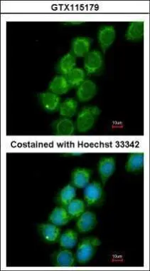
PRC1 antibody [N1N2], N-termGTX115179
ApplicationsImmunoFluorescence, Western Blot, ImmunoCytoChemistry, ImmunoHistoChemistry, ImmunoHistoChemistry Paraffin
ReactivityHuman, Mouse
TargetPRC1
- SizePrice

Product group Antibodies
Anti-PRC1 AntibodyHPA034521
ApplicationsWestern Blot, ImmunoHistoChemistry
ReactivityHuman, Mouse, Rat
TargetPRC1
- SizePrice
Product group Antibodies
Anti-PRC1 AntibodyCAB7029
ApplicationsImmunoFluorescence, Western Blot, ELISA, ImmunoCytoChemistry, ImmunoHistoChemistry, ImmunoHistoChemistry Paraffin
ReactivityHuman
TargetPRC1
- SizePrice