


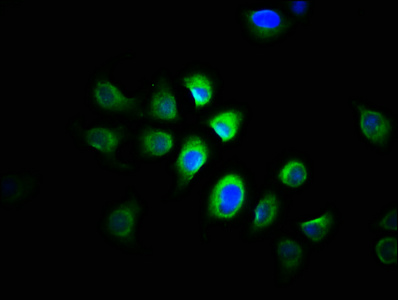
Immunofluorescent staining of human cell line A549 shows localization to nucleoplasm.

Immunofluorescent staining of human cell line A549 shows localization to nucleoplasm.
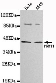
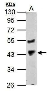
Anti-PRMT1 Antibody
HPA072136
ApplicationsWestern Blot, ImmunoCytoChemistry
Product group Antibodies
ReactivityHuman
TargetPRMT1
Overview
- SupplierAtlas Antibodies
- Product NameAnti-PRMT1 Antibody
- Delivery Days Customer4
- ApplicationsWestern Blot, ImmunoCytoChemistry
- CertificationResearch Use Only
- ClonalityPolyclonal
- ConjugateUnconjugated
- Gene ID3276
- Target namePRMT1
- Target descriptionprotein arginine methyltransferase 1
- Target synonymsANM1, HCP1, HRMT1L2, IR1B4, protein arginine N-methyltransferase 1, HMT1 (hnRNP methyltransferase, S. cerevisiae)-like 2, heterogeneous nuclear ribonucleoprotein methyltransferase 1-like 2, highly conserved protein 1, histone-arginine N-methyltransferase PRMT1, interferon receptor 1-bound protein 4
- HostRabbit
- IsotypeIgG
- Scientific DescriptionRecombinant Protein Epitope Signature Tag (PrEST) antigen sequence
- ReactivityHuman
- Storage Instruction-20°C,2°C to 8°C
- UNSPSC41116161
Datasheet
MSDS
Related products
Product group Antibodies
Anti-PRMT1 Antibody144-61934
ApplicationsImmunoFluorescence, Western Blot, ImmunoHistoChemistry
ReactivityHuman, Mouse, Rat
TargetPRMT1
- SizePrice

Product group Antibodies
Anti-PRMT1 Antibody Picoband(r)A01417-3-CARRIER-FREE
ApplicationsFlow Cytometry, Western Blot, ELISA, ImmunoHistoChemistry
ReactivityHuman
TargetPRMT1
- SizePrice
Product group Antibodies
PRMT1 Recombinant Antibody, Biotin ConjugatedBSM-61453R-BIOTIN
ApplicationsImmunoPrecipitation, Western Blot, ImmunoHistoChemistry, ImmunoHistoChemistry Frozen, ImmunoHistoChemistry Paraffin
ReactivityHuman, Mouse, Rat
TargetPRMT1
- SizePrice
Product group Antibodies
PRMT1 Polyclonal AntibodyCAC14032
ApplicationsImmunoFluorescence, Western Blot, ELISA, ImmunoHistoChemistry
ReactivityMouse
TargetPRMT1
- SizePrice
Product group Antibodies
PRMT1 AntibodyCSB-PA03025A0RB
ApplicationsImmunoFluorescence, Western Blot, ELISA, ImmunoHistoChemistry
ReactivityHuman, Mouse
TargetPRMT1
- SizePrice
Product group Antibodies

PRMT1 AntibodyLS-C331224
ApplicationsWestern Blot, ImmunoHistoChemistry
ReactivityHuman, Mouse, Rat
TargetPRMT1
- SizePrice
Product group Antibodies
PRMT1 antibodyGTX128211
ApplicationsImmunoFluorescence, Western Blot, ImmunoCytoChemistry
ReactivityHuman, Zebra Fish
TargetPRMT1
- SizePrice