


Immunohistochemical staining of human duodenum shows strong cytoplasmic and membranous positivity in glandular cells.

Immunohistochemical staining of human duodenum shows strong cytoplasmic and membranous positivity in glandular cells.
Anti-PRMT7 Antibody
HPA044241
ApplicationsImmunoCytoChemistry, ImmunoHistoChemistry
Product group Antibodies
ReactivityHuman
TargetPRMT7
Overview
- SupplierAtlas Antibodies
- Product NameAnti-PRMT7 Antibody
- Delivery Days Customer4
- ApplicationsImmunoCytoChemistry, ImmunoHistoChemistry
- CertificationResearch Use Only
- ClonalityPolyclonal
- ConjugateUnconjugated
- Gene ID54496
- Target namePRMT7
- Target descriptionprotein arginine methyltransferase 7
- Target synonymsSBIDDS, protein arginine N-methyltransferase 7, [Myelin basic protein]-arginine N-methyltransferase PRMT7, histone-arginine N-methyltransferase PRMT7
- HostRabbit
- IsotypeIgG
- Protein IDQ9NVM4
- Protein NameProtein arginine N-methyltransferase 7
- Scientific DescriptionRecombinant Protein Epitope Signature Tag (PrEST) antigen sequence
- ReactivityHuman
- Storage Instruction-20°C,2°C to 8°C
- UNSPSC41116161
Datasheet
MSDS
Related products
Product group Antibodies
Anti-PRMT7 AntibodyA80946
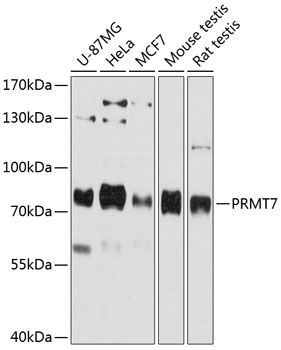
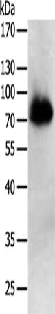
ApplicationsWestern Blot
ReactivityHuman, Mouse, Rat
- SizePrice

Product group Antibodies
Anti-PRMT7 Antibody Picoband(r)A05485-2-CARRIER-FREE
ApplicationsFlow Cytometry, ImmunoFluorescence, Western Blot, ELISA, ImmunoCytoChemistry, ImmunoHistoChemistry
ReactivityHuman, Mouse, Rat
TargetPRMT7
- SizePrice
Product group Antibodies
Anti-PRMT7 Antibody144-12159
ApplicationsWestern Blot
ReactivityHuman, Mouse, Rat
TargetPRMT7
- SizePrice

Product group Antibodies
ApplicationsFlow Cytometry, Western Blot, ImmunoCytoChemistry
ReactivityHuman, Mouse, Rat
TargetPRMT7
- SizePrice
Product group Antibodies

Goat anti-PRMT7EB11563
ApplicationsWestern Blot, ELISA
ReactivityCanine, Human, Mouse, Rat
TargetPRMT7
- SizePrice
Product group Antibodies
Prmt7 Polyclonal AntibodyCAC07835
ApplicationsImmunoFluorescence, Western Blot, ELISA, ImmunoHistoChemistry
ReactivityMouse
TargetPRMT7
- SizePrice
Product group Antibodies
PRMT7 AntibodyCSB-PA036451
ApplicationsWestern Blot, ELISA
ReactivityHuman
TargetPRMT7
- SizePrice
Product group Antibodies

PRMT7 AntibodyLS-C400983
ApplicationsWestern Blot, ELISA
ReactivityHuman
TargetPRMT7
- SizePrice