


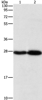
Figure 1. Western blot analysis of Proteasome 20S Beta 7/PSMB7 using anti-Proteasome 20S Beta 7/PSMB7 antibody (A08095-1). Electrophoresis was performed on a 5-20% SDS-PAGE gel at 70V (Stacking gel) / 90V (Resolving gel) for 2-3 hours. The sample well of each lane was loaded with 30 ug of sample under reducing conditions. Lane 1: human Hela whole cell lysates, Lane 2: human PC-3 whole cell lysates, Lane 3: human MCF-7 whole cell lysates, Lane 4: human Caco-2 whole cell lysates, Lane 5: human 293T whole cell lysates, Lane 6; human HepG2 whole cell lysates, Lane 7: human HL-60 whole cell lysates, Lane 8: human A549 whole cell lysates. After electrophoresis, proteins were transferred to a nitrocellulose membrane at 150 mA for 50-90 minutes. Blocked the membrane with 5% non-fat milk/TBS for 1.5 hour at RT. The membrane was incubated with rabbit anti-Proteasome 20S Beta 7/PSMB7 antigen affinity purified polyclonal antibody (Catalog # A08095-1) at 0.5 microg/mL overnight at 4°C, then washed with TBS-0.1%Tween 3 times with 5 minutes each and probed with a goat anti-rabbit IgG-HRP secondary antibody at a dilution of 1:5000 for 1.5 hour at RT. The signal is developed using an Enhanced Chemiluminescent detection (ECL) kit (Catalog # EK1002) with Tanon 5200 system. A specific band was detected for Proteasome 20S Beta 7/PSMB7 at approximately 26 kDa. The expected band size for Proteasome 20S Beta 7/PSMB7 is at 26 kDa.
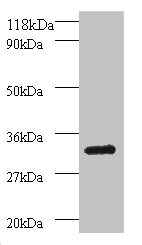
. Electrophoresis was performed on a 5-20% SDS-PAGE gel at 70V (Stacking gel) / 90V (Resolving gel) for 2-3 hours. The sample well of each lane was loaded with 30 ug of sample under reducing conditions. Lane 1: rat brain tissue lysates, Lane 2: rat liver tissue lysates, Lane 3: rat kidney tissue lysates, Lane 4: rat pancreas tissue lysates, Lane 5: mouse brain tissue lysates, Lane 6; mouse liver tissue lysates, Lane 7: mouse kidney tissue lysates. After electrophoresis, proteins were transferred to a nitrocellulose membrane at 150 mA for 50-90 minutes. Blocked the membrane with 5% non-fat milk/TBS for 1.5 hour at RT. The membrane was incubated with rabbit anti-Proteasome 20S Beta 7/PSMB7 antigen affinity purified polyclonal antibody (Catalog # A08095-1) at 0.5 microg/mL overnight at 4°C, then washed with TBS-0.1%Tween 3 times with 5 minutes each and probed with a goat anti-rabbit IgG-HRP secondary antibody at a dilution of 1:5000 for 1.5 hour at RT. The signal is developed using an Enhanced Chemiluminescent detection (ECL) kit (Catalog # EK1002) with Tanon 5200 system. A specific band was detected for Proteasome 20S Beta 7/PSMB7 at approximately 26 kDa. The expected band size for Proteasome 20S Beta 7/PSMB7 is at 26 kDa.")
. Proteasome 20S Beta 7/PSMB7 was detected in a paraffin-embedded section of mouse brain tissue. Heat mediated antigen retrieval was performed in EDTA buffer (pH 8.0, epitope retrieval solution). The tissue section was blocked with 10% goat serum. The tissue section was then incubated with 2 microg/ml rabbit anti-Proteasome 20S Beta 7/PSMB7 Antibody (A08095-1) overnight at 4°C. Peroxidase Conjugated Goat Anti-rabbit IgG was used as secondary antibody and incubated for 30 minutes at 37°C. The tissue section was developed using HRP Conjugated Rabbit IgG Super Vision Assay Kit (Catalog # SV0002) with DAB as the chromogen.")
. Proteasome 20S Beta 7/PSMB7 was detected in a paraffin-embedded section of rat brain tissue. Heat mediated antigen retrieval was performed in EDTA buffer (pH 8.0, epitope retrieval solution). The tissue section was blocked with 10% goat serum. The tissue section was then incubated with 2 microg/ml rabbit anti-Proteasome 20S Beta 7/PSMB7 Antibody (A08095-1) overnight at 4°C. Peroxidase Conjugated Goat Anti-rabbit IgG was used as secondary antibody and incubated for 30 minutes at 37°C. The tissue section was developed using HRP Conjugated Rabbit IgG Super Vision Assay Kit (Catalog # SV0002) with DAB as the chromogen.")
. Proteasome 20S Beta 7/PSMB7 was detected in a paraffin-embedded section of human renal clear cell carcinoma tissue. Heat mediated antigen retrieval was performed in EDTA buffer (pH 8.0, epitope retrieval solution). The tissue section was blocked with 10% goat serum. The tissue section was then incubated with 2 microg/ml rabbit anti-Proteasome 20S Beta 7/PSMB7 Antibody (A08095-1) overnight at 4°C. Peroxidase Conjugated Goat Anti-rabbit IgG was used as secondary antibody and incubated for 30 minutes at 37°C. The tissue section was developed using HRP Conjugated Rabbit IgG Super Vision Assay Kit (Catalog # SV0002) with DAB as the chromogen.")
. Proteasome 20S Beta 7/PSMB7 was detected in a paraffin-embedded section of human cervical intraepithelial neoplasia tissue. Heat mediated antigen retrieval was performed in EDTA buffer (pH 8.0, epitope retrieval solution). The tissue section was blocked with 10% goat serum. The tissue section was then incubated with 2 microg/ml rabbit anti-Proteasome 20S Beta 7/PSMB7 Antibody (A08095-1) overnight at 4°C. Peroxidase Conjugated Goat Anti-rabbit IgG was used as secondary antibody and incubated for 30 minutes at 37°C. The tissue section was developed using HRP Conjugated Rabbit IgG Super Vision Assay Kit (Catalog # SV0002) with DAB as the chromogen.")
. Proteasome 20S Beta 7/PSMB7 was detected in a paraffin-embedded section of human colorectal cancer tissue. Heat mediated antigen retrieval was performed in EDTA buffer (pH 8.0, epitope retrieval solution). The tissue section was blocked with 10% goat serum. The tissue section was then incubated with 2 microg/ml rabbit anti-Proteasome 20S Beta 7/PSMB7 Antibody (A08095-1) overnight at 4°C. Peroxidase Conjugated Goat Anti-rabbit IgG was used as secondary antibody and incubated for 30 minutes at 37°C. The tissue section was developed using HRP Conjugated Rabbit IgG Super Vision Assay Kit (Catalog # SV0002) with DAB as the chromogen.")
. Proteasome 20S Beta 7/PSMB7 was detected in a paraffin-embedded section of human invasive breast carcinoma tissue. Heat mediated antigen retrieval was performed in EDTA buffer (pH 8.0, epitope retrieval solution). The tissue section was blocked with 10% goat serum. The tissue section was then incubated with 2 microg/ml rabbit anti-Proteasome 20S Beta 7/PSMB7 Antibody (A08095-1) overnight at 4°C. Peroxidase Conjugated Goat Anti-rabbit IgG was used as secondary antibody and incubated for 30 minutes at 37°C. The tissue section was developed using HRP Conjugated Rabbit IgG Super Vision Assay Kit (Catalog # SV0002) with DAB as the chromogen.")
. Proteasome 20S Beta 7/PSMB7 was detected in a paraffin-embedded section of human liver cancer tissue. Heat mediated antigen retrieval was performed in EDTA buffer (pH 8.0, epitope retrieval solution). The tissue section was blocked with 10% goat serum. The tissue section was then incubated with 2 microg/ml rabbit anti-Proteasome 20S Beta 7/PSMB7 Antibody (A08095-1) overnight at 4°C. Peroxidase Conjugated Goat Anti-rabbit IgG was used as secondary antibody and incubated for 30 minutes at 37°C. The tissue section was developed using HRP Conjugated Rabbit IgG Super Vision Assay Kit (Catalog # SV0002) with DAB as the chromogen.")
. Proteasome 20S Beta 7/PSMB7 was detected in a paraffin-embedded section of human ovarian serous adenocarcinoma tissue. Heat mediated antigen retrieval was performed in EDTA buffer (pH 8.0, epitope retrieval solution). The tissue section was blocked with 10% goat serum. The tissue section was then incubated with 2 microg/ml rabbit anti-Proteasome 20S Beta 7/PSMB7 Antibody (A08095-1) overnight at 4°C. Peroxidase Conjugated Goat Anti-rabbit IgG was used as secondary antibody and incubated for 30 minutes at 37°C. The tissue section was developed using HRP Conjugated Rabbit IgG Super Vision Assay Kit (Catalog # SV0002) with DAB as the chromogen.")
Figure 1. Western blot analysis of Proteasome 20S Beta 7/PSMB7 using anti-Proteasome 20S Beta 7/PSMB7 antibody (A08095-1). Electrophoresis was performed on a 5-20% SDS-PAGE gel at 70V (Stacking gel) / 90V (Resolving gel) for 2-3 hours. The sample well of each lane was loaded with 30 ug of sample under reducing conditions. Lane 1: human Hela whole cell lysates, Lane 2: human PC-3 whole cell lysates, Lane 3: human MCF-7 whole cell lysates, Lane 4: human Caco-2 whole cell lysates, Lane 5: human 293T whole cell lysates, Lane 6; human HepG2 whole cell lysates, Lane 7: human HL-60 whole cell lysates, Lane 8: human A549 whole cell lysates. After electrophoresis, proteins were transferred to a nitrocellulose membrane at 150 mA for 50-90 minutes. Blocked the membrane with 5% non-fat milk/TBS for 1.5 hour at RT. The membrane was incubated with rabbit anti-Proteasome 20S Beta 7/PSMB7 antigen affinity purified polyclonal antibody (Catalog # A08095-1) at 0.5 microg/mL overnight at 4°C, then washed with TBS-0.1%Tween 3 times with 5 minutes each and probed with a goat anti-rabbit IgG-HRP secondary antibody at a dilution of 1:5000 for 1.5 hour at RT. The signal is developed using an Enhanced Chemiluminescent detection (ECL) kit (Catalog # EK1002) with Tanon 5200 system. A specific band was detected for Proteasome 20S Beta 7/PSMB7 at approximately 26 kDa. The expected band size for Proteasome 20S Beta 7/PSMB7 is at 26 kDa.
Anti-Proteasome 20S beta 7/PSMB7 Antibody Picoband(r)
A08095-1-CARRIER-FREE
ApplicationsFlow Cytometry, ImmunoFluorescence, Western Blot, ELISA, ImmunoCytoChemistry, ImmunoHistoChemistry
Product group Antibodies
ReactivityHuman, Mouse, Rat
TargetPSMB7
Overview
- SupplierBoster Bio
- Product NameAnti-Proteasome 20S beta 7/PSMB7 Antibody Picoband(r)
- Delivery Days Customer9
- ApplicationsFlow Cytometry, ImmunoFluorescence, Western Blot, ELISA, ImmunoCytoChemistry, ImmunoHistoChemistry
- CertificationResearch Use Only
- ClonalityPolyclonal
- Concentration500 ug/ml
- Gene ID5695
- Target namePSMB7
- Target descriptionproteasome 20S subunit beta 7
- Target synonymsproteasome subunit beta type-7, beta-2, epididymis secretory sperm binding protein, macropain chain Z, multicatalytic endopeptidase complex chain Z, proteasome (prosome, macropain) subunit, beta type, 7, proteasome catalytic subunit 2, proteasome subunit Z, proteasome subunit alpha, proteasome subunit beta 7, proteasome subunit beta-2, proteasome subunit beta2, protein serine kinase c17
- HostRabbit
- IsotypeIgG
- Protein IDQ99436
- Protein NameProteasome subunit beta type-7
- Scientific DescriptionBoster Bio Anti-Proteasome 20S beta 7/PSMB7 Antibody Picoband® catalog # A08095-1. Tested in ELISA, Flow Cytometry, IF, IHC, ICC, WB applications. This antibody reacts with Human, Mouse, Rat. The brand Picoband indicates this is a premium antibody that guarantees superior quality, high affinity, and strong signals with minimal background in Western blot applications. Only our best-performing antibodies are designated as Picoband, ensuring unmatched performance.
- ReactivityHuman, Mouse, Rat
- Storage Instruction-20°C,2°C to 8°C
- UNSPSC12352203
Related products
Product group Antibodies
Anti-PSMB7 Antibody144-60785
ApplicationsWestern Blot
ReactivityHuman, Mouse, Rat
TargetPSMB7
- SizePrice
Product group Antibodies
PSMB7 Polyclonal AntibodyCAC13818
ApplicationsImmunoFluorescence, Western Blot, ELISA, ImmunoHistoChemistry
TargetPSMB7
- SizePrice
Product group Antibodies

PSMB7 AntibodyLS-C749754
ApplicationsWestern Blot
ReactivityHuman, Mouse, Rat
TargetPSMB7
- SizePrice
Product group Antibodies
Anti-PSMB7 AntibodyA37875
ApplicationsWestern Blot, ImmunoHistoChemistry
ReactivityHuman, Mouse
- SizePrice
Product group Antibodies
PSMB7 AntibodyCSB-PA01345A0RB
ApplicationsImmunoFluorescence, Western Blot, ELISA, ImmunoHistoChemistry
ReactivityHuman
TargetPSMB7
- SizePrice

Product group Antibodies
Anti-PSMB7 AntibodyHPA052408
ApplicationsImmunoHistoChemistry
ReactivityHuman
TargetPSMB7
- SizePrice
Product group Antibodies

Goat anti-PSMB7 AntibodyEB10298
ApplicationsWestern Blot, ELISA
ReactivityBovine, Human, Mouse, Porcine, Rat
TargetPSMB7
- SizePrice
Product group Antibodies
References
ApplicationsImmunoFluorescence, Western Blot, ImmunoCytoChemistry, ImmunoHistoChemistry, ImmunoHistoChemistry Paraffin
ReactivityHuman, Mouse
TargetPSMB7
- SizePrice