

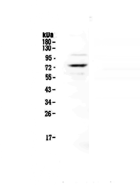
Figure 1. Western blot analysis of Protein S using anti-Protein S antibody (A01568-1). Electrophoresis was performed on a 5-20% SDS-PAGE gel at 70V (Stacking gel) / 90V (Resolving gel) for 2-3 hours. The sample well of each lane was loaded with 50ug of sample under reducing conditions. Lane 1: human HepG2 whole cell lysates. After Electrophoresis, proteins were transferred to a Nitrocellulose membrane at 150mA for 50-90 minutes. Blocked the membrane with 5% Non-fat Milk/ TBS for 1.5 hour at RT. The membrane was incubated with rabbit anti-Protein S antigen affinity purified polyclonal antibody (Catalog # A01568-1) at 0.5 microg/mL overnight at 4°C, then washed with TBS-0.1%Tween 3 times with 5 minutes each and probed with a goat anti-rabbit IgG-HRP secondary antibody at a dilution of 1:10000 for 1.5 hour at RT. The signal is developed using an Enhanced Chemiluminescent detection (ECL) kit (Catalog # EK1002) with Tanon 5200 system. A specific band was detected for Protein S at approximately 75KD. The expected band size for Protein S is at 75KD.
Figure 1. Western blot analysis of Protein S using anti-Protein S antibody (A01568-1). Electrophoresis was performed on a 5-20% SDS-PAGE gel at 70V (Stacking gel) / 90V (Resolving gel) for 2-3 hours. The sample well of each lane was loaded with 50ug of sample under reducing conditions. Lane 1: human HepG2 whole cell lysates. After Electrophoresis, proteins were transferred to a Nitrocellulose membrane at 150mA for 50-90 minutes. Blocked the membrane with 5% Non-fat Milk/ TBS for 1.5 hour at RT. The membrane was incubated with rabbit anti-Protein S antigen affinity purified polyclonal antibody (Catalog # A01568-1) at 0.5 microg/mL overnight at 4°C, then washed with TBS-0.1%Tween 3 times with 5 minutes each and probed with a goat anti-rabbit IgG-HRP secondary antibody at a dilution of 1:10000 for 1.5 hour at RT. The signal is developed using an Enhanced Chemiluminescent detection (ECL) kit (Catalog # EK1002) with Tanon 5200 system. A specific band was detected for Protein S at approximately 75KD. The expected band size for Protein S is at 75KD.
Anti-Protein S/PROS1 Antibody Picoband(r)
A01568-1-CARRIER-FREE
ApplicationsWestern Blot, ELISA
Product group Antibodies
ReactivityHuman
TargetPROS1
Overview
- SupplierBoster Bio
- Product NameAnti-Protein S/PROS1 Antibody Picoband(r)
- Delivery Days Customer9
- ApplicationsWestern Blot, ELISA
- CertificationResearch Use Only
- ClonalityPolyclonal
- Concentration500 ug/ml
- Gene ID5627
- Target namePROS1
- Target descriptionprotein S
- Target synonymsPROS, PS21, PS22, PS23, PS24, PS25, PSA, THPH5, THPH6, vitamin K-dependent protein S, protein S (alpha), protein Sa, vitamin K-dependent plasma protein S
- HostRabbit
- IsotypeIgG
- Protein IDP07225
- Protein NameVitamin K-dependent protein S
- Scientific DescriptionBoster Bio Anti-Protein S/PROS1 Antibody Picoband® catalog # A01568-1. Tested in ELISA, WB applications. This antibody reacts with Human. The brand Picoband indicates this is a premium antibody that guarantees superior quality, high affinity, and strong signals with minimal background in Western blot applications. Only our best-performing antibodies are designated as Picoband, ensuring unmatched performance.
- ReactivityHuman
- Storage Instruction-20°C,2°C to 8°C
- UNSPSC12352203
Related products
Product group Antibodies
PROS1 AntibodyCSB-PA018754LA01HU
ApplicationsImmunoFluorescence, ELISA, ImmunoHistoChemistry
ReactivityHuman
TargetPROS1
- SizePrice

Product group Antibodies
Anti-PROS1 AntibodyA29963
ApplicationsWestern Blot, ImmunoHistoChemistry
ReactivityHuman, Mouse, Rat
- SizePrice

Product group Antibodies
Anti-PROS1 AntibodyHPA007724
ApplicationsWestern Blot, ImmunoHistoChemistry
ReactivityHuman
TargetPROS1
- SizePrice

Product group Antibodies

PROS1 / Protein S AntibodyLS-C401125
ApplicationsELISA, ImmunoHistoChemistry
ReactivityHuman
TargetPROS1
- SizePrice
Product group Antibodies
ApplicationsImmunoPrecipitation, Western Blot, ImmunoCytoChemistry, ImmunoHistoChemistry
ReactivityRat
TargetPROS1
- SizePrice
Product group Antibodies
Protein S antibodyGTX66404
ApplicationsWestern Blot
ReactivityHuman, Mouse, Rat
TargetPROS1
- SizePrice
Product group Antibodies
Protein S Polyclonal AntibodyBS-9512R
ApplicationsImmunoFluorescence, Western Blot, ImmunoHistoChemistry, ImmunoHistoChemistry Frozen, ImmunoHistoChemistry Paraffin
ReactivityHuman, Mouse, Rat
TargetPROS1
- SizePrice