


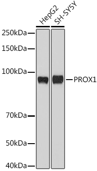
Western blot analysis of PROX1 expression in HepG2 cell lysate.
Western blot analysis of PROX1 expression in HepG2 cell lysate.
Anti-PROX1 Rabbit Monoclonal Antibody
M01985-1
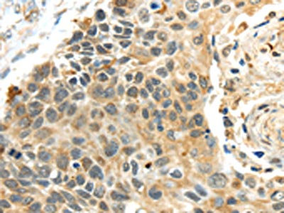
ApplicationsWestern Blot, ImmunoHistoChemistry
Product group Antibodies
ReactivityHuman
TargetPROX1
Overview
- SupplierBoster Bio
- Product NameAnti-PROX1 Rabbit Monoclonal Antibody
- Delivery Days Customer9
- ApplicationsWestern Blot, ImmunoHistoChemistry
- CertificationResearch Use Only
- ClonalityMonoclonal
- Clone IDAOGA-16
- Gene ID5629
- Target namePROX1
- Target descriptionprospero homeobox 1
- Target synonymsprospero homeobox protein 1, homeobox prospero-like protein PROX1, prospero-related homeobox 1
- HostRabbit
- IsotypeIgG
- Protein IDQ92786
- Protein NameProspero homeobox protein 1
- Scientific DescriptionBoster Bio Anti-PROX1 Rabbit Monoclonal Antibody catalog # M01985-1. Tested in IHC, WB application. This antibody reacts with Human.
- ReactivityHuman
- Storage Instruction-20°C
- UNSPSC12352203
Datasheet
MSDS
Related products
Product group Antibodies
Anti-PROX1 AntibodyA91318
ApplicationsWestern Blot
ReactivityHuman, Mouse, Rat
- SizePrice
Product group Antibodies
anti-PROX1 (human), pAbAG-25T-0104
ApplicationsWestern Blot, ImmunoHistoChemistry
ReactivityHuman
TargetPROX1
- SizePrice
Product group Antibodies
Anti-PROX1 Antibody144-62605
ApplicationsWestern Blot
ReactivityHuman, Mouse, Rat
TargetPROX1
- SizePrice

Product group Antibodies
Anti-PROX1 AntibodyAMAB91863
ApplicationsWestern Blot, ImmunoHistoChemistry
ReactivityHuman, Mouse, Rat
TargetPROX1
- SizePrice
Product group Antibodies

PROX1 AntibodyLS-C748105
ApplicationsWestern Blot
ReactivityHuman
TargetPROX1
- SizePrice

Product group Antibodies
Anti-PROX1 Antibody Picoband(r)A01985-1-CARRIER-FREE
ApplicationsFlow Cytometry, ImmunoFluorescence, ImmunoPrecipitation, Western Blot, ELISA, ImmunoCytoChemistry
ReactivityHuman, Rat
TargetPROX1
- SizePrice
Product group Antibodies
References
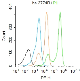
Prox1 Polyclonal AntibodyBS-2774R
ApplicationsFlow Cytometry, ImmunoFluorescence, Western Blot, ELISA, ImmunoHistoChemistry, ImmunoHistoChemistry Paraffin
ReactivityBovine, Canine, Equine, Human, Mouse, Porcine, Rabbit, Rat
TargetPROX1
- SizePrice
Product group Antibodies
PROX1 Polyclonal AntibodyCAC15141
ApplicationsWestern Blot, ELISA
TargetPROX1
- SizePrice
Product group Antibodies
PROX1 AntibodyCSB-PA215840
ApplicationsWestern Blot, ELISA, ImmunoHistoChemistry
ReactivityHuman, Mouse
TargetPROX1
- SizePrice