


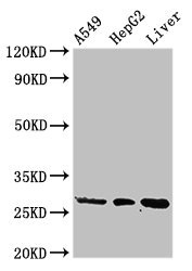
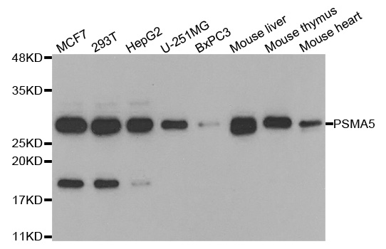
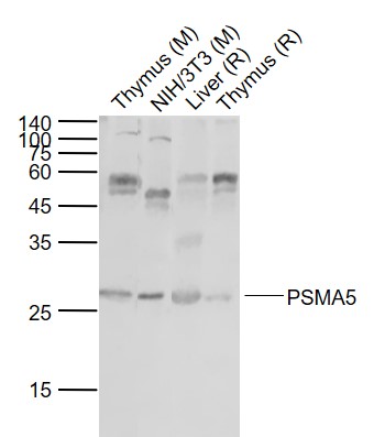
Figure 1. Western blot analysis of PSMA5 using anti-PSMA5 antibody (A07499-1). Electrophoresis was performed on a 5-20% SDS-PAGE gel at 70V (Stacking gel) / 90V (Resolving gel) for 2-3 hours. The sample well of each lane was loaded with 30 ug of sample under reducing conditions. Lane 1: human Hela whole cell lysates, Lane 2: human HepG2 whole cell lysates, Lane 3: human 293T whole cell lysates, Lane 4: monkey COS-7 whole cell lysates, Lane 5: rat brain tissue lysates, Lane 6: rat PC-12 whole cell lysates, Lane 7: mouse brain tissue lysates, Lane 8: mouse Neuro-2a whole cell lysates. After electrophoresis, proteins were transferred to a nitrocellulose membrane at 150 mA for 50-90 minutes. Blocked the membrane with 5% non-fat milk/TBS for 1.5 hour at RT. The membrane was incubated with rabbit anti-PSMA5 antigen affinity purified polyclonal antibody (Catalog # A07499-1) at 0.5 microg/mL overnight at 4°C, then washed with TBS-0.1%Tween 3 times with 5 minutes each and probed with a goat anti-rabbit IgG-HRP secondary antibody at a dilution of 1:5000 for 1.5 hour at RT. The signal is developed using an Enhanced Chemiluminescent detection (ECL) kit (Catalog # EK1002) with Tanon 5200 system. A specific band was detected for PSMA5 at approximately 26 kDa. The expected band size for PSMA5 is at 26 kDa.
. PSMA5 was detected in an immunocytochemical section of U2OS cells. Enzyme antigen retrieval was performed using IHC enzyme antigen retrieval reagent (AR0022) for 15 mins. The cells were blocked with 10% goat serum. And then incubated with 5 microg/mL rabbit anti-PSMA5 Antibody (A07499-1) overnight at 4°C. DyLight®488 Conjugated Goat Anti-Rabbit IgG (BA1127) was used as secondary antibody at 1:500 dilution and incubated for 30 minutes at 37°C. The section was counterstained with DAPI. Visualize using a fluorescence microscope and filter sets appropriate for the label used.")
. Overlay histogram showing Hela cells stained with A07499-1 (Blue line). To facilitate intracellular staining, cells were fixed with 4% paraformaldehyde and permeabilized with permeabilization buffer. The cells were blocked with 10% normal goat serum. And then incubated with rabbit anti-PSMA5 Antibody (A07499-1, 1 microg/1x106 cells) for 30 min at 20°C. DyLight®488 conjugated goat anti-rabbit IgG (BA1127, 5-10 microg/1x106 cells) was used as secondary antibody for 30 minutes at 20°C. Isotype control antibody (Green line) was rabbit IgG (1 microg/1x106) used under the same conditions. Unlabelled sample (Red line) was also used as a control.")
Figure 1. Western blot analysis of PSMA5 using anti-PSMA5 antibody (A07499-1). Electrophoresis was performed on a 5-20% SDS-PAGE gel at 70V (Stacking gel) / 90V (Resolving gel) for 2-3 hours. The sample well of each lane was loaded with 30 ug of sample under reducing conditions. Lane 1: human Hela whole cell lysates, Lane 2: human HepG2 whole cell lysates, Lane 3: human 293T whole cell lysates, Lane 4: monkey COS-7 whole cell lysates, Lane 5: rat brain tissue lysates, Lane 6: rat PC-12 whole cell lysates, Lane 7: mouse brain tissue lysates, Lane 8: mouse Neuro-2a whole cell lysates. After electrophoresis, proteins were transferred to a nitrocellulose membrane at 150 mA for 50-90 minutes. Blocked the membrane with 5% non-fat milk/TBS for 1.5 hour at RT. The membrane was incubated with rabbit anti-PSMA5 antigen affinity purified polyclonal antibody (Catalog # A07499-1) at 0.5 microg/mL overnight at 4°C, then washed with TBS-0.1%Tween 3 times with 5 minutes each and probed with a goat anti-rabbit IgG-HRP secondary antibody at a dilution of 1:5000 for 1.5 hour at RT. The signal is developed using an Enhanced Chemiluminescent detection (ECL) kit (Catalog # EK1002) with Tanon 5200 system. A specific band was detected for PSMA5 at approximately 26 kDa. The expected band size for PSMA5 is at 26 kDa.
Anti-PSMA5 Antibody Picoband(r)
A07499-1-CARRIER-FREE
ApplicationsFlow Cytometry, ImmunoFluorescence, Western Blot, ELISA, ImmunoCytoChemistry
Product group Antibodies
ReactivityHuman, Monkey, Mouse, Rat
TargetPSMA5
Overview
- SupplierBoster Bio
- Product NameAnti-PSMA5 Antibody Picoband(r)
- Delivery Days Customer9
- ApplicationsFlow Cytometry, ImmunoFluorescence, Western Blot, ELISA, ImmunoCytoChemistry
- CertificationResearch Use Only
- ClonalityPolyclonal
- Concentration500 ug/ml
- Gene ID5686
- Target namePSMA5
- Target descriptionproteasome 20S subunit alpha 5
- Target synonymsPSC5, ZETA, proteasome subunit alpha type-5, alpha-5, epididymis tissue sperm binding protein Li 11n, macropain subunit zeta, macropain zeta chain, multicatalytic endopeptidase complex zeta chain, proteasome (prosome, macropain) subunit, alpha type, 5, proteasome alpha 5 subunit, proteasome component 5, proteasome subunit alpha 5, proteasome subunit zeta, proteasome zeta chain
- HostRabbit
- Protein IDP28066
- Protein NameProteasome subunit alpha type-5
- Scientific DescriptionBoster Bio Anti-PSMA5 Antibody Picoband® catalog # A07499-1. Tested in WB, ICC/IF, Flow Cytometry, ELISA applications. This antibody reacts with Human, Monkey, Mouse, Rat. The brand Picoband indicates this is a premium antibody that guarantees superior quality, high affinity, and strong signals with minimal background in Western blot applications. Only our best-performing antibodies are designated as Picoband, ensuring unmatched performance.
- ReactivityHuman, Monkey, Mouse, Rat
- Storage Instruction-20°C,2°C to 8°C
- UNSPSC12352203
Related products
Product group Antibodies
PSMA5 AntibodyCSB-PA018870LA01HU
ApplicationsWestern Blot, ELISA, ImmunoHistoChemistry
ReactivityHuman, Rat
TargetPSMA5
- SizePrice
Product group Antibodies
Anti-PSMA5 AntibodyA31027
ApplicationsImmunoFluorescence, Western Blot, ImmunoHistoChemistry
ReactivityHuman, Mouse, Rat
- SizePrice
Product group Antibodies

PSMA5 AntibodyLS-C749070
ApplicationsWestern Blot, ImmunoHistoChemistry
ReactivityHuman, Mouse, Rat
TargetPSMA5
- SizePrice
Product group Antibodies
Anti-PSMA5 AntibodyHPA028392
ApplicationsWestern Blot, ImmunoHistoChemistry
ReactivityHuman
TargetPSMA5
- SizePrice
Product group Antibodies
PSMA5 Polyclonal AntibodyCAC14872
ApplicationsWestern Blot, ELISA, ImmunoHistoChemistry
ReactivityRat
TargetPSMA5
- SizePrice
Product group Antibodies
PSMA5 antibodyGTX103845
ApplicationsImmunoFluorescence, Western Blot, ImmunoCytoChemistry, ImmunoHistoChemistry, ImmunoHistoChemistry Paraffin
ReactivityHuman, Mouse
TargetPSMA5
- SizePrice
Product group Antibodies
Anti-PSMA5 Antibody144-05752
ApplicationsImmunoFluorescence, Western Blot
ReactivityHuman, Mouse
TargetPSMA5
- SizePrice
Product group Antibodies
PSMA5 Polyclonal AntibodyBS-9354R
ApplicationsImmunoFluorescence, Western Blot, ImmunoHistoChemistry, ImmunoHistoChemistry Paraffin
ReactivityBovine, Chicken, Human, Mouse, Porcine, Rabbit, Rat
TargetPSMA5
- SizePrice