


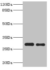
Figure 1. Western blot analysis of PSME1 using anti-PSME1 antibody (A04638-2). Electrophoresis was performed on a 5-20% SDS-PAGE gel at 70V (Stacking gel) / 90V (Resolving gel) for 2-3 hours. The sample well of each lane was loaded with 30 ug of sample under reducing conditions. Lane 1: human Raji whole cell lysates, Lane 2: human A431 whole cell lysates, Lane 3: rat liver tissue lysates, Lane 4: rat spleen tissue lysates, Lane 5: rat RH35 whole cell lysates, Lane 6: mouse liver tissue lysates, Lane 7: mouse spleen tissue lysates, Lane 8: mouse RAW264.7 whole cell lysates. After electrophoresis, proteins were transferred to a nitrocellulose membrane at 150 mA for 50-90 minutes. Blocked the membrane with 5% non-fat milk/TBS for 1.5 hour at RT. The membrane was incubated with rabbit anti-PSME1 antigen affinity purified polyclonal antibody (Catalog # A04638-2) at 0.25 microg/mL overnight at 4°C, then washed with TBS-0.1%Tween 3 times with 5 minutes each and probed with a goat anti-rabbit IgG-HRP secondary antibody at a dilution of 1:5000 for 1.5 hour at RT. The signal is developed using an Enhanced Chemiluminescent detection (ECL) kit (Catalog # EK1002) with Tanon 5200 system. A specific band was detected for PSME1 at approximately 30 kDa. The expected band size for PSME1 is at 29 kDa.
. PSME1 was detected in an immunocytochemical section of A549 cells. Enzyme antigen retrieval was performed using IHC enzyme antigen retrieval reagent (AR0022) for 15 mins. The cells were blocked with 10% goat serum. And then incubated with 5 microg/mL rabbit anti-PSME1 Antibody (A04638-2) overnight at 4°C. Cy3 Conjugated Goat Anti-Rabbit IgG (BA1032) was used as secondary antibody at 1:500 dilution and incubated for 30 minutes at 37°C. The section was counterstained with DAPI. Visualize using a fluorescence microscope and filter sets appropriate for the label used.")
. Overlay histogram showing JK cells stained with A04638-2 (Blue line). To facilitate intracellular staining, cells were fixed with 4% paraformaldehyde and permeabilized with permeabilization buffer. The cells were blocked with 10% normal goat serum. And then incubated with rabbit anti-PSME1 Antibody (A04638-2, 1 microg/1x106 cells) for 30 min at 20°C. DyLight®488 conjugated goat anti-rabbit IgG (BA1127, 5-10 microg/1x106 cells) was used as secondary antibody for 30 minutes at 20°C. Isotype control antibody (Green line) was rabbit IgG (1 microg/1x106) used under the same conditions. Unlabelled sample (Red line) was also used as a control.")
Figure 1. Western blot analysis of PSME1 using anti-PSME1 antibody (A04638-2). Electrophoresis was performed on a 5-20% SDS-PAGE gel at 70V (Stacking gel) / 90V (Resolving gel) for 2-3 hours. The sample well of each lane was loaded with 30 ug of sample under reducing conditions. Lane 1: human Raji whole cell lysates, Lane 2: human A431 whole cell lysates, Lane 3: rat liver tissue lysates, Lane 4: rat spleen tissue lysates, Lane 5: rat RH35 whole cell lysates, Lane 6: mouse liver tissue lysates, Lane 7: mouse spleen tissue lysates, Lane 8: mouse RAW264.7 whole cell lysates. After electrophoresis, proteins were transferred to a nitrocellulose membrane at 150 mA for 50-90 minutes. Blocked the membrane with 5% non-fat milk/TBS for 1.5 hour at RT. The membrane was incubated with rabbit anti-PSME1 antigen affinity purified polyclonal antibody (Catalog # A04638-2) at 0.25 microg/mL overnight at 4°C, then washed with TBS-0.1%Tween 3 times with 5 minutes each and probed with a goat anti-rabbit IgG-HRP secondary antibody at a dilution of 1:5000 for 1.5 hour at RT. The signal is developed using an Enhanced Chemiluminescent detection (ECL) kit (Catalog # EK1002) with Tanon 5200 system. A specific band was detected for PSME1 at approximately 30 kDa. The expected band size for PSME1 is at 29 kDa.
Anti-PSME1 Antibody Picoband(r)
A04638-2-CARRIER-FREE
ApplicationsFlow Cytometry, ImmunoFluorescence, Western Blot, ELISA, ImmunoCytoChemistry
Product group Antibodies
ReactivityHuman, Mouse, Rat
TargetPSME1
Overview
- SupplierBoster Bio
- Product NameAnti-PSME1 Antibody Picoband(r)
- Delivery Days Customer9
- ApplicationsFlow Cytometry, ImmunoFluorescence, Western Blot, ELISA, ImmunoCytoChemistry
- CertificationResearch Use Only
- ClonalityPolyclonal
- Concentration500 ug/ml
- Gene ID5720
- Target namePSME1
- Target descriptionproteasome activator subunit 1
- Target synonymsHEL-S-129m, IFI5111, PA28A, PA28alpha, REGalpha, proteasome activator complex subunit 1, 11S regulator complex subunit alpha, 29-kD MCP activator subunit, IGUP I-5111, activator of multicatalytic protease subunit 1, epididymis secretory sperm binding protein Li 129m, interferon gamma up-regulated I-5111 protein, interferon-gamma IEF SSP 5111, interferon-gamma-inducible protein 5111, proteasome (prosome, macropain) activator subunit 1 (PA28 alpha)
- HostRabbit
- IsotypeIgG
- Protein IDQ06323
- Protein NameProteasome activator complex subunit 1
- Scientific DescriptionBoster Bio Anti-PSME1 Antibody Picoband® catalog # A04638-2. Tested in ELISA, Flow Cytometry, IF, ICC, WB applications. This antibody reacts with Human, Mouse, Rat. The brand Picoband indicates this is a premium antibody that guarantees superior quality, high affinity, and strong signals with minimal background in Western blot applications. Only our best-performing antibodies are designated as Picoband, ensuring unmatched performance.
- ReactivityHuman, Mouse, Rat
- Storage Instruction-20°C,2°C to 8°C
- UNSPSC12352203
Related products
Product group Antibodies
PSME1 AntibodyCSB-PA018915DSR1HU
ApplicationsImmunoPrecipitation, Western Blot, ELISA, ImmunoHistoChemistry
ReactivityHuman, Mouse
TargetPSME1
- SizePrice
Product group Antibodies
Anti-PSME1 AntibodyA30753
ApplicationsImmunoFluorescence, Western Blot, ImmunoHistoChemistry
ReactivityHuman, Mouse, Rat
- SizePrice
Product group Antibodies

PSME1 AntibodyLS-C830948
ApplicationsELISA, ImmunoHistoChemistry
ReactivityHuman, Mouse, Rat
TargetPSME1
- SizePrice
Product group Antibodies

Goat anti-PSME1 (isoform 1)EB07761
ApplicationsWestern Blot, ELISA
ReactivityCanine, Human, Mouse, Rat
TargetPSME1
- SizePrice

Product group Antibodies
Anti-PSME1 AntibodyHPA006632
ApplicationsWestern Blot, ImmunoHistoChemistry
ReactivityHuman
TargetPSME1
- SizePrice
Product group Antibodies
PSME1 Recombinant Antibody, AbBy Fluor-555 ConjugatedBSM-61868R-BF555
ApplicationsWestern Blot
ReactivityHuman, Mouse, Rat
TargetPSME1
- SizePrice
Product group Antibodies
Psme1 Recombinant AntibodyCAC12533
ApplicationsFlow Cytometry, ImmunoFluorescence, Western Blot, ELISA, ImmunoHistoChemistry
TargetPSME1
- SizePrice
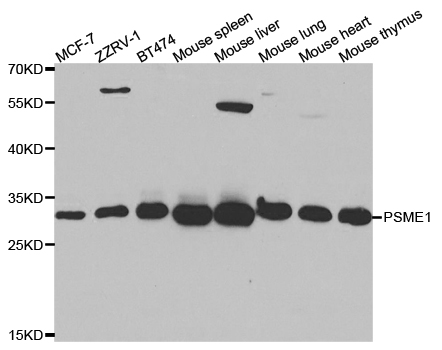
![Various whole cell extracts (30 μg) were separated by 12% SDS-PAGE, and the membrane was blotted with PSME1 antibody [N1C3] (GTX104422) diluted at 1:5000. The HRP-conjugated anti-rabbit IgG antibody (GTX213110-01) was used to detect the primary antibody. Corresponding RNA expression data for the same cell lines are based on Human Protein Atlas program.](https://www.genetex.com/upload/website/prouct_img/normal/GTX104422/GTX104422_40639_20240329_WB_TPM_watermark_24040123_413.webp)
Product group Antibodies
PSME1 antibody [N1C3]GTX104422
ApplicationsWestern Blot
ReactivityHuman
TargetPSME1
- SizePrice
Product group Antibodies
Anti-PSME1 Antibody144-05358
ApplicationsImmunoFluorescence, Western Blot, ImmunoHistoChemistry
ReactivityHuman, Mouse, Rat
TargetPSME1
- SizePrice