


Immunohistochemical staining of human stomach shows moderate nuclear positivity in glandular cells.

Immunohistochemical staining of human stomach shows moderate nuclear positivity in glandular cells.
Anti-PYCRL Antibody
HPA063416
ApplicationsImmunoCytoChemistry, ImmunoHistoChemistry
Product group Antibodies
ReactivityHuman
TargetPYCR3
Overview
- SupplierAtlas Antibodies
- Product NameAnti-PYCRL Antibody
- Delivery Days Customer4
- ApplicationsImmunoCytoChemistry, ImmunoHistoChemistry
- CertificationResearch Use Only
- ClonalityPolyclonal
- ConjugateUnconjugated
- Gene ID65263
- Target namePYCR3
- Target descriptionpyrroline-5-carboxylate reductase 3
- Target synonymsPYCRL, pyrroline-5-carboxylate reductase 3, P5C reductase 3, P5CR 3, pyrroline-5-carboxylate reductase-like
- HostRabbit
- IsotypeIgG
- Protein IDQ53H96
- Protein NamePyrroline-5-carboxylate reductase 3
- Scientific DescriptionRecombinant Protein Epitope Signature Tag (PrEST) antigen sequence
- ReactivityHuman
- Storage Instruction-20°C,2°C to 8°C
- UNSPSC41116161
Datasheet
MSDS
Related products

Product group Antibodies
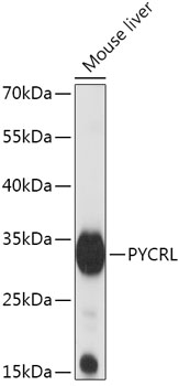
Anti-PYCRL/PYCR3 Antibody Picoband(r)A32428-1-CARRIER-FREE
ApplicationsFlow Cytometry, Western Blot, ELISA
ReactivityHuman, Mouse
TargetPYCR3
- SizePrice

Product group Antibodies
Anti-PYCRL AntibodyHPA069706
ApplicationsImmunoHistoChemistry
ReactivityHuman
TargetPYCR3
- SizePrice
Product group Antibodies

PYCRL Antibody (aa221-255)LS-C356651
ApplicationsWestern Blot, ELISA
ReactivityHuman
TargetPYCR3
- SizePrice
![WB analysis of HEK293T cells transfected with PYCRL plasmid (Right) or empty vector (Left) for 48 hrs using GTX83750 PYCRL antibody [2E10]. Loading : 5 ug per lane](https://www.genetex.com/upload/website/prouct_img/normal/GTX83750/GTX83750_3877_WB_w_23061420_861.webp)
Product group Antibodies
PYCRL antibody [2E10]GTX83750
ApplicationsFlow Cytometry, ImmunoFluorescence, Western Blot, ImmunoCytoChemistry
ReactivityHuman, Monkey
TargetPYCR3
- SizePrice
Product group Antibodies
Anti-PYCRL Antibody144-65144
ApplicationsWestern Blot
ReactivityHuman, Mouse
TargetPYCR3
- SizePrice