


Figure 1. Western blot analysis of Pyrin/MEFV using anti-Pyrin/MEFV antibody (A00764-1). Electrophoresis was performed on a 5-20% SDS-PAGE gel at 70V (Stacking gel) / 90V (Resolving gel) for 2-3 hours. The sample well of each lane was loaded with 30 ug of sample under reducing conditions. Lane 1: human Hela whole cell lysates, Lane 2: human U-87MG whole cell lysates, Lane 3: human Raji whole cell lysates, Lane 4: mouse spleen tissue lysates, Lane 5: mouse lung tissue lysates. After electrophoresis, proteins were transferred to a nitrocellulose membrane at 150 mA for 50-90 minutes. Blocked the membrane with 5% non-fat milk/TBS for 1.5 hour at RT. The membrane was incubated with rabbit anti-Pyrin/MEFV antigen affinity purified polyclonal antibody (Catalog # A00764-1) at 0.5 microg/mL overnight at 4°C, then washed with TBS-0.1%Tween 3 times with 5 minutes each and probed with a goat anti-rabbit IgG-HRP secondary antibody at a dilution of 1:5000 for 1.5 hour at RT. The signal is developed using an Enhanced Chemiluminescent detection (ECL) kit (Catalog # EK1002) with Tanon 5200 system. A specific band was detected for Pyrin/MEFV at approximately 86 kDa. The expected band size for Pyrin/MEFV is at 86 kDa.
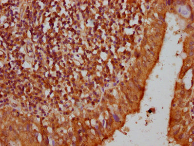
. Pyrin/MEFV was detected in a paraffin-embedded section of human thyroid papillary carcinoma tissue. Heat mediated antigen retrieval was performed in EDTA buffer (pH 8.0, epitope retrieval solution). The tissue section was blocked with 10% goat serum. The tissue section was then incubated with 2 microg/ml rabbit anti-Pyrin/MEFV Antibody (A00764-1) overnight at 4°C. Biotinylated goat anti-rabbit IgG was used as secondary antibody and incubated for 30 minutes at 37°C. The tissue section was developed using Strepavidin-Biotin-Complex (SABC) (Catalog # SA1022) with DAB as the chromogen.")
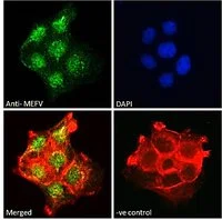
. Pyrin/MEFV was detected in an immunocytochemical section of Hela cells. Enzyme antigen retrieval was performed using IHC enzyme antigen retrieval reagent (AR0022) for 15 mins. The cells were blocked with 10% goat serum. And then incubated with 5 microg/mL rabbit anti-Pyrin/MEFV Antibody (A00764-1) overnight at 4°C. DyLight®488 Conjugated Goat Anti-Rabbit IgG (BA1127) was used as secondary antibody at 1:100 dilution and incubated for 30 minutes at 37°C. The section was counterstained with DAPI. Visualize using a fluorescence microscope and filter sets appropriate for the label used.")
. Overlay histogram showing U937 cells stained with A00764-1 (Blue line). To facilitate intracellular staining, cells were fixed with 4% paraformaldehyde and permeabilized with permeabilization buffer. The cells were blocked with 10% normal goat serum. And then incubated with rabbit anti-Pyrin/MEFV Antibody (A00764-1, 1 microg/1x106 cells) for 30 min at 20°C. DyLight®488 conjugated goat anti-rabbit IgG (BA1127, 5-10 microg/1x106 cells) was used as secondary antibody for 30 minutes at 20°C. Isotype control antibody (Green line) was rabbit IgG (1 microg/1x106) used under the same conditions. Unlabelled sample without incubation with primary antibody and secondary antibody (Red line) was used as a blank control.")
Figure 1. Western blot analysis of Pyrin/MEFV using anti-Pyrin/MEFV antibody (A00764-1). Electrophoresis was performed on a 5-20% SDS-PAGE gel at 70V (Stacking gel) / 90V (Resolving gel) for 2-3 hours. The sample well of each lane was loaded with 30 ug of sample under reducing conditions. Lane 1: human Hela whole cell lysates, Lane 2: human U-87MG whole cell lysates, Lane 3: human Raji whole cell lysates, Lane 4: mouse spleen tissue lysates, Lane 5: mouse lung tissue lysates. After electrophoresis, proteins were transferred to a nitrocellulose membrane at 150 mA for 50-90 minutes. Blocked the membrane with 5% non-fat milk/TBS for 1.5 hour at RT. The membrane was incubated with rabbit anti-Pyrin/MEFV antigen affinity purified polyclonal antibody (Catalog # A00764-1) at 0.5 microg/mL overnight at 4°C, then washed with TBS-0.1%Tween 3 times with 5 minutes each and probed with a goat anti-rabbit IgG-HRP secondary antibody at a dilution of 1:5000 for 1.5 hour at RT. The signal is developed using an Enhanced Chemiluminescent detection (ECL) kit (Catalog # EK1002) with Tanon 5200 system. A specific band was detected for Pyrin/MEFV at approximately 86 kDa. The expected band size for Pyrin/MEFV is at 86 kDa.
Anti-Pyrin/MEFV Antibody Picoband(r)
A00764-1-CARRIER-FREE
ApplicationsFlow Cytometry, ImmunoFluorescence, Western Blot, ELISA, ImmunoCytoChemistry, ImmunoHistoChemistry
Product group Antibodies
ReactivityHuman, Mouse
TargetMEFV
Overview
- SupplierBoster Bio
- Product NameAnti-Pyrin/MEFV Antibody Picoband(r)
- Delivery Days Customer9
- ApplicationsFlow Cytometry, ImmunoFluorescence, Western Blot, ELISA, ImmunoCytoChemistry, ImmunoHistoChemistry
- CertificationResearch Use Only
- ClonalityPolyclonal
- Concentration500 ug/ml
- Gene ID4210
- Target nameMEFV
- Target descriptionMEFV innate immunity regulator, pyrin
- Target synonymsFMF, MEF, PAAND, TRIM20, pyrin, MEFV innate immuity regulator, pyrin, MEFV, pyrin innate immunity regulator, Mediterranean fever, marenostrin
- HostRabbit
- IsotypeIgG
- Protein IDO15553
- Protein NamePyrin
- Scientific DescriptionBoster Bio Anti-Pyrin/MEFV Antibody Picoband® catalog # A00764-1. Tested in ELISA, Flow Cytometry, IF, IHC, ICC, WB applications. This antibody reacts with Human, Mouse. The brand Picoband indicates this is a premium antibody that guarantees superior quality, high affinity, and strong signals with minimal background in Western blot applications. Only our best-performing antibodies are designated as Picoband, ensuring unmatched performance.
- ReactivityHuman, Mouse
- Storage Instruction-20°C,2°C to 8°C
- UNSPSC12352203
Related products
Product group Antibodies
MEFV AntibodyCSB-PA013675LA01HU
ApplicationsELISA, ImmunoHistoChemistry
ReactivityHuman
TargetMEFV
- SizePrice
Product group Antibodies

Goat anti-MEFVEB06877
ApplicationsFlow Cytometry, ImmunoFluorescence, ELISA
ReactivityHuman
TargetMEFV
- SizePrice

Product group Antibodies
Anti-MEFV AntibodyHPA077497
ApplicationsImmunoCytoChemistry
ReactivityHuman
TargetMEFV
- SizePrice
Product group Antibodies

Pyrin / MEFV / MEF AntibodyLS-C411133
ApplicationsWestern Blot
ReactivityHuman
TargetMEFV
- SizePrice
Product group Antibodies
MEFV antibodyGTX01092
ApplicationsFlow Cytometry, ImmunoFluorescence, ImmunoCytoChemistry
ReactivityHuman
TargetMEFV
- SizePrice