

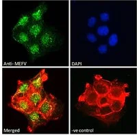
ICC/IF analysis of PFA-fixed A431 cells using GTX01092 MEFV antibody. Green : Primary antibody Red : Actin Blue : DAPI Permeabilization : 0.15% Triton X-100 Dilution : 10μg/ml

ICC/IF analysis of PFA-fixed A431 cells using GTX01092 MEFV antibody. Green : Primary antibody Red : Actin Blue : DAPI Permeabilization : 0.15% Triton X-100 Dilution : 10μg/ml
MEFV antibody
GTX01092
ApplicationsFlow Cytometry, ImmunoFluorescence, ImmunoCytoChemistry
Product group Antibodies
ReactivityHuman
TargetMEFV
Overview
- SupplierGeneTex
- Product NameMEFV antibody
- Delivery Days Customer9
- Application Supplier NoteICC/IF: 10microg/ml. FCM: 10microg/ml. *Optimal dilutions/concentrations should be determined by the researcher.Not tested in other applications.
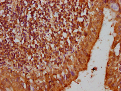
- ApplicationsFlow Cytometry, ImmunoFluorescence, ImmunoCytoChemistry
- CertificationResearch Use Only
- ClonalityPolyclonal
- Concentration0.5 mg/ml
- ConjugateUnconjugated
- Gene ID4210
- Target nameMEFV
- Target descriptionMEFV innate immunity regulator, pyrin
- Target synonymsFMF, MEF, PAAND, TRIM20, pyrin, MEFV innate immuity regulator, pyrin, MEFV, pyrin innate immunity regulator, Mediterranean fever, marenostrin
- HostGoat
- IsotypeIgG
- Protein IDO15553
- Protein NamePyrin
- Scientific DescriptionThis gene encodes a protein, also known as pyrin or marenostrin, that is an important modulator of innate immunity. Mutations in this gene are associated with Mediterranean fever, a hereditary periodic fever syndrome. [provided by RefSeq, Jul 2008]
- ReactivityHuman
- Storage Instruction-20°C or -80°C,2°C to 8°C
- UNSPSC41116161
Datasheet
Related products
Product group Antibodies
MEFV AntibodyCSB-PA013675LA01HU
ApplicationsELISA, ImmunoHistoChemistry
ReactivityHuman
TargetMEFV
- SizePrice

Product group Antibodies
Anti-Pyrin/MEFV Antibody Picoband(r)A00764-1-CARRIER-FREE
ApplicationsFlow Cytometry, ImmunoFluorescence, Western Blot, ELISA, ImmunoCytoChemistry, ImmunoHistoChemistry
ReactivityHuman, Mouse
TargetMEFV
- SizePrice
Product group Antibodies

Goat anti-MEFVEB06877
ApplicationsFlow Cytometry, ImmunoFluorescence, ELISA
ReactivityHuman
TargetMEFV
- SizePrice

Product group Antibodies
Anti-MEFV AntibodyHPA077497
ApplicationsImmunoCytoChemistry
ReactivityHuman
TargetMEFV
- SizePrice
Product group Antibodies

Pyrin / MEFV / MEF AntibodyLS-C411133
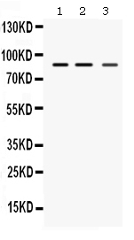
ApplicationsWestern Blot
ReactivityHuman
TargetMEFV
- SizePrice
Product group Antibodies
MEFV antibodyGTX89483
ApplicationsWestern Blot
ReactivityHuman
TargetMEFV
- SizePrice
Product group Antibodies
MEFV antibodyGTX01088
ApplicationsWestern Blot
ReactivityHuman, Rat
TargetMEFV
- SizePrice