


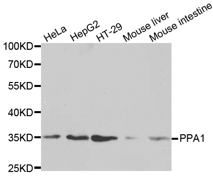
Figure 1. Western blot analysis of PPA1 using anti-PPA1 antibody (A07485-1). Electrophoresis was performed on a 5-20% SDS-PAGE gel at 70V (Stacking gel) / 90V (Resolving gel) for 2-3 hours. The sample well of each lane was loaded with 30 ug of sample under reducing conditions. Lane 1: human U937 whole cell lysates, Lane 2: human CACO-2 whole cell lysates, Lane 3: human Raji whole cell lysates, Lane 4: human PC-3 whole cell lysates, Lane 5: human HEK293 whole cell lysates, Lane 6: human HL-60 whole cell lysates, Lane 7: human K562 whole cell lysates, Lane 8: rat liver tissue lysates, Lane 9: mouse liver tissue lysates. After electrophoresis, proteins were transferred to a nitrocellulose membrane at 150 mA for 50-90 minutes. Blocked the membrane with 5% non-fat milk/TBS for 1.5 hour at RT. The membrane was incubated with rabbit anti-PPA1 antigen affinity purified polyclonal antibody (Catalog # A07485-1) at 0.5 microg/mL overnight at 4°C, then washed with TBS-0.1%Tween 3 times with 5 minutes each and probed with a goat anti-rabbit IgG-HRP secondary antibody at a dilution of 1:5000 for 1.5 hour at RT. The signal is developed using an Enhanced Chemiluminescent detection (ECL) kit (Catalog # EK1002) with Tanon 5200 system. A specific band was detected for PPA1 at approximately 33 kDa. The expected band size for PPA1 is at 33 kDa.
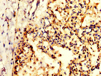
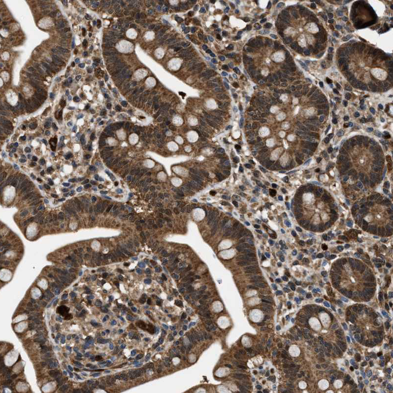
. PPA1 was detected in paraffin-embedded section of human intestinal cancer tissue. Heat mediated antigen retrieval was performed in citrate buffer (pH6, epitope retrieval solution) for 20 mins. The tissue section was blocked with 10% goat serum. The tissue section was then incubated with 1microg/ml rabbit anti-PPA1 Antibody (A07485-1) overnight at 4°C. Biotinylated goat anti-rabbit IgG was used as secondary antibody and incubated for 30 minutes at 37°C. The tissue section was developed using Strepavidin-Biotin-Complex (SABC)(Catalog # SA1022) with DAB as the chromogen.")
. PPA1 was detected in paraffin-embedded section of mouse brain tissue. Heat mediated antigen retrieval was performed in citrate buffer (pH6, epitope retrieval solution) for 20 mins. The tissue section was blocked with 10% goat serum. The tissue section was then incubated with 1microg/ml rabbit anti-PPA1 Antibody (A07485-1) overnight at 4°C. Biotinylated goat anti-rabbit IgG was used as secondary antibody and incubated for 30 minutes at 37°C. The tissue section was developed using Strepavidin-Biotin-Complex (SABC)(Catalog # SA1022) with DAB as the chromogen.")
. PPA1 was detected in paraffin-embedded section of human lung cancer tissue. Heat mediated antigen retrieval was performed in citrate buffer (pH6, epitope retrieval solution) for 20 mins. The tissue section was blocked with 10% goat serum. The tissue section was then incubated with 1microg/ml rabbit anti-PPA1 Antibody (A07485-1) overnight at 4°C. Biotinylated goat anti-rabbit IgG was used as secondary antibody and incubated for 30 minutes at 37°C. The tissue section was developed using Strepavidin-Biotin-Complex (SABC)(Catalog # SA1022) with DAB as the chromogen.")
. PPA1 was detected in paraffin-embedded section of human mammary cancer tissue. Heat mediated antigen retrieval was performed in citrate buffer (pH6, epitope retrieval solution) for 20 mins. The tissue section was blocked with 10% goat serum. The tissue section was then incubated with 1microg/ml rabbit anti-PPA1 Antibody (A07485-1) overnight at 4°C. Biotinylated goat anti-rabbit IgG was used as secondary antibody and incubated for 30 minutes at 37°C. The tissue section was developed using Strepavidin-Biotin-Complex (SABC)(Catalog # SA1022) with DAB as the chromogen.")
. PPA1 was detected in paraffin-embedded section of rat brain tissue. Heat mediated antigen retrieval was performed in citrate buffer (pH6, epitope retrieval solution) for 20 mins. The tissue section was blocked with 10% goat serum. The tissue section was then incubated with 1microg/ml rabbit anti-PPA1 Antibody (A07485-1) overnight at 4°C. Biotinylated goat anti-rabbit IgG was used as secondary antibody and incubated for 30 minutes at 37°C. The tissue section was developed using Strepavidin-Biotin-Complex (SABC)(Catalog # SA1022) with DAB as the chromogen.")
Figure 1. Western blot analysis of PPA1 using anti-PPA1 antibody (A07485-1). Electrophoresis was performed on a 5-20% SDS-PAGE gel at 70V (Stacking gel) / 90V (Resolving gel) for 2-3 hours. The sample well of each lane was loaded with 30 ug of sample under reducing conditions. Lane 1: human U937 whole cell lysates, Lane 2: human CACO-2 whole cell lysates, Lane 3: human Raji whole cell lysates, Lane 4: human PC-3 whole cell lysates, Lane 5: human HEK293 whole cell lysates, Lane 6: human HL-60 whole cell lysates, Lane 7: human K562 whole cell lysates, Lane 8: rat liver tissue lysates, Lane 9: mouse liver tissue lysates. After electrophoresis, proteins were transferred to a nitrocellulose membrane at 150 mA for 50-90 minutes. Blocked the membrane with 5% non-fat milk/TBS for 1.5 hour at RT. The membrane was incubated with rabbit anti-PPA1 antigen affinity purified polyclonal antibody (Catalog # A07485-1) at 0.5 microg/mL overnight at 4°C, then washed with TBS-0.1%Tween 3 times with 5 minutes each and probed with a goat anti-rabbit IgG-HRP secondary antibody at a dilution of 1:5000 for 1.5 hour at RT. The signal is developed using an Enhanced Chemiluminescent detection (ECL) kit (Catalog # EK1002) with Tanon 5200 system. A specific band was detected for PPA1 at approximately 33 kDa. The expected band size for PPA1 is at 33 kDa.
Anti-Pyrophosphatase 1/PPA1 Antibody Picoband(r)
A07485-1-CARRIER-FREE
ApplicationsWestern Blot, ELISA, ImmunoHistoChemistry
Product group Antibodies
ReactivityHuman, Mouse, Rat
TargetPPA1
Overview
- SupplierBoster Bio
- Product NameAnti-Pyrophosphatase 1/PPA1 Antibody Picoband(r)
- Delivery Days Customer9
- ApplicationsWestern Blot, ELISA, ImmunoHistoChemistry
- CertificationResearch Use Only
- ClonalityPolyclonal
- Concentration500 ug/ml
- Gene ID5464
- Target namePPA1
- Target descriptioninorganic pyrophosphatase 1
- Target synonymsHEL-S-66p, IOPPP, PP, PP1, SID6-8061, inorganic pyrophosphatase, PPase, cytosolic inorganic pyrophosphatase, diphosphate phosphohydrolase, epididymis secretory sperm binding protein Li 66p, inorganic diphosphatase 1, pyrophosphatase (inorganic) 1, pyrophosphatase 1, pyrophosphate phospho-hydrolase
- HostRabbit
- IsotypeIgG
- Protein IDQ15181
- Protein NameInorganic pyrophosphatase
- Scientific DescriptionBoster Bio Anti-Pyrophosphatase 1/PPA1 Antibody Picoband® catalog # A07485-1. Tested in ELISA, IHC, WB applications. This antibody reacts with Human, Mouse, Rat. The brand Picoband indicates this is a premium antibody that guarantees superior quality, high affinity, and strong signals with minimal background in Western blot applications. Only our best-performing antibodies are designated as Picoband, ensuring unmatched performance.
- ReactivityHuman, Mouse, Rat
- Storage Instruction-20°C,2°C to 8°C
- UNSPSC12352203
Related products
Product group Antibodies
PPA1 Monoclonal AntibodyCAC13709
ApplicationsFlow Cytometry, ImmunoFluorescence, Western Blot, ELISA, ImmunoHistoChemistry
TargetPPA1
- SizePrice
Product group Antibodies
PPA1 AntibodyCSB-PA614884LA01HU
ApplicationsImmunoFluorescence, ELISA, ImmunoHistoChemistry
ReactivityHuman
TargetPPA1
- SizePrice
Product group Antibodies
Anti-PPA1 Antibody144-02108
ApplicationsWestern Blot, ImmunoHistoChemistry
ReactivityHuman, Mouse, Rat
TargetPPA1
- SizePrice
Product group Antibodies
Anti-PPA1 AntibodyA29952
ApplicationsWestern Blot, ImmunoHistoChemistry
ReactivityHuman, Mouse, Rat
- SizePrice
Product group Antibodies

PPA1 AntibodyLS-C749062
ApplicationsWestern Blot
ReactivityHuman, Mouse, Rat
TargetPPA1
- SizePrice
Product group Antibodies
Anti-PPA1 AntibodyHPA019878
ApplicationsWestern Blot, ImmunoHistoChemistry
ReactivityHuman
TargetPPA1
- SizePrice
Product group Antibodies
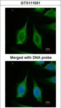
PPA1 antibodyGTX111031
ApplicationsImmunoFluorescence, Western Blot, ImmunoCytoChemistry, ImmunoHistoChemistry, ImmunoHistoChemistry Paraffin
ReactivityHuman, Mouse, Rat
TargetPPA1
- SizePrice
Product group Antibodies
Anti-PPA1Y158351
ApplicationsWestern Blot, ELISA, ImmunoHistoChemistry
ReactivityHuman
- SizePrice