


Anti-RAB34 Antibody
A100645
ApplicationsELISA, ImmunoHistoChemistry
Product group Antibodies
ReactivityHuman
Overview
- SupplierAntibodies.com
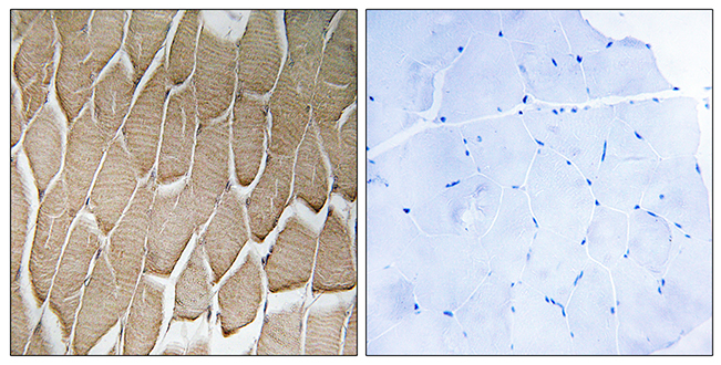
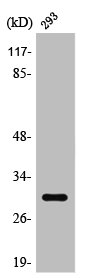
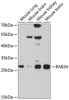
- Product NameAnti-RAB34 Antibody
- Delivery Days Customer7
- ApplicationsELISA, ImmunoHistoChemistry
- CertificationResearch Use Only
- ClonalityPolyclonal
- ConjugateUnconjugated
- HostRabbit
- IsotypeIgG
- Scientific DescriptionRabbit polyclonal antibody to RAB34.
- ReactivityHuman
- UNSPSC12352203
Related products
Product group Antibodies
RAB34 AntibodyCSB-PA003895
ApplicationsWestern Blot, ELISA, ImmunoHistoChemistry
ReactivityHuman
TargetRAB34
- SizePrice

Product group Antibodies
Anti-RAB34 Antibody Picoband(r)A11097-3-CARRIER-FREE
ApplicationsFlow Cytometry, Western Blot, ELISA
ReactivityHuman, Mouse, Rat
TargetRAB34
- SizePrice
Product group Antibodies
Anti-RAB34 Antibody144-10332
ApplicationsWestern Blot
ReactivityHuman, Mouse, Rat
TargetRAB34
- SizePrice

Product group Antibodies
Anti-RAB34 AntibodyHPA021366
ApplicationsImmunoHistoChemistry
ReactivityHuman
TargetRAB34
- SizePrice
Product group Antibodies

RAB34 Antibody (C-Terminus)LS-C368783
ApplicationsWestern Blot, ImmunoHistoChemistry, ImmunoHistoChemistry Paraffin
ReactivityHuman
TargetRAB34
- SizePrice
Product group Antibodies
RAB34 antibodyGTX65558
ApplicationsWestern Blot
ReactivityHuman, Mouse
TargetRAB34
- SizePrice