

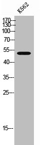
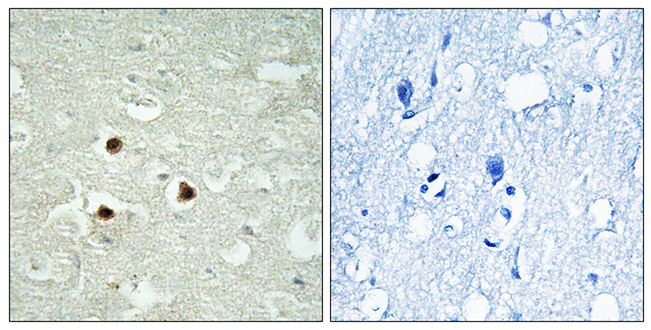
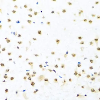
Anti-RAB3IP Antibody
144-08094
ApplicationsWestern Blot, ImmunoHistoChemistry
Product group Antibodies
ReactivityHuman, Mouse, Rat
TargetRAB3IP
Overview
- SupplierRayBiotech
- Product NameAnti-RAB3IP Antibody
- Delivery Days Customer16
- ApplicationsWestern Blot, ImmunoHistoChemistry
- CertificationResearch Use Only
- ClonalityPolyclonal
- ConjugateUnconjugated
- Gene ID117177
- Target nameRAB3IP
- Target descriptionRAB3A interacting protein
- Target synonymsRABIN3, RABIN8, rab-3A-interacting protein, SSX2 interacting protein, rab3A-interacting protein, rabin-3
- HostRabbit
- IsotypeIgG
- Protein IDQ96QF0
- Protein NameRab-3A-interacting protein
- Scientific DescriptionRAB3IP Polyclonal Antibody
- ReactivityHuman, Mouse, Rat
- Storage Instruction-20°C
- UNSPSC12352203
Related products
Product group Antibodies
RAB3IP AntibodyCSB-PA006887
ApplicationsWestern Blot, ELISA, ImmunoHistoChemistry
ReactivityHuman, Mouse, Rat
TargetRAB3IP
- SizePrice

Product group Antibodies
Anti-RAB3IP Antibody Picoband(r)A09085-3-CARRIER-FREE
ApplicationsFlow Cytometry, Western Blot, ELISA, ImmunoHistoChemistry
ReactivityHuman, Mouse, Rat
TargetRAB3IP
- SizePrice
Product group Antibodies
Anti-RAB3IP AntibodyA96624
ApplicationsELISA, ImmunoHistoChemistry
ReactivityHuman, Mouse, Rat
- SizePrice
Product group Antibodies

RAB3IP / RABIN3 AntibodyLS-C830952
ApplicationsELISA, ImmunoHistoChemistry
ReactivityHuman, Mouse, Rat
TargetRAB3IP
- SizePrice

Product group Antibodies
Anti-RAB3IP AntibodyHPA039794
ApplicationsWestern Blot, ImmunoCytoChemistry, ImmunoHistoChemistry
ReactivityHuman
TargetRAB3IP
- SizePrice
Product group Antibodies
RAB3IP antibodyGTX55771
ApplicationsWestern Blot, ImmunoHistoChemistry, ImmunoHistoChemistry Paraffin
ReactivityHuman, Mouse, Rat
TargetRAB3IP
- SizePrice