


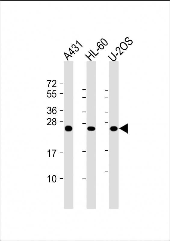
Figure 1. Western blot analysis of RAB5C/RABL using anti-RAB5C/RABL antibody (M05148-1). Electrophoresis was performed on a 5-20% SDS-PAGE gel at 70V (Stacking gel) / 90V (Resolving gel) for 2-3 hours. The sample well of each lane was loaded with 30 ug of sample under reducing conditions. Lane 1: human 293T whole cell lysates, Lane 2: human HepG2 whole cell lysates, Lane 3: human A549 whole cell lysates, Lane 4: human RT4 whole cell lysates, Lane 5: rat RH35 whole cell lysates, Lane 6: mouse HEPA1-6 whole cell lysates. After electrophoresis, proteins were transferred to a nitrocellulose membrane at 150 mA for 50-90 minutes. Blocked the membrane with 5% non-fat milk/TBS for 1.5 hour at RT. The membrane was incubated with rabbit anti-RAB5C/RABL antigen affinity purified monoclonal antibody (Catalog # M05148-1) at 1:500 overnight at 4°C, then washed with TBS-0.1%Tween 3 times with 5 minutes each and probed with a goat anti-rabbit IgG-HRP secondary antibody at a dilution of 1:1000 for 1.5 hour at RT. The signal is developed using an Enhanced Chemiluminescent detection (ECL) kit (Catalog # EK1002) with Tanon 5200 system. A specific band was detected for RAB5C/RABL at approximately 23 kDa. The expected band size for RAB5C/RABL is at 23 kDa.
Figure 1. Western blot analysis of RAB5C/RABL using anti-RAB5C/RABL antibody (M05148-1). Electrophoresis was performed on a 5-20% SDS-PAGE gel at 70V (Stacking gel) / 90V (Resolving gel) for 2-3 hours. The sample well of each lane was loaded with 30 ug of sample under reducing conditions. Lane 1: human 293T whole cell lysates, Lane 2: human HepG2 whole cell lysates, Lane 3: human A549 whole cell lysates, Lane 4: human RT4 whole cell lysates, Lane 5: rat RH35 whole cell lysates, Lane 6: mouse HEPA1-6 whole cell lysates. After electrophoresis, proteins were transferred to a nitrocellulose membrane at 150 mA for 50-90 minutes. Blocked the membrane with 5% non-fat milk/TBS for 1.5 hour at RT. The membrane was incubated with rabbit anti-RAB5C/RABL antigen affinity purified monoclonal antibody (Catalog # M05148-1) at 1:500 overnight at 4°C, then washed with TBS-0.1%Tween 3 times with 5 minutes each and probed with a goat anti-rabbit IgG-HRP secondary antibody at a dilution of 1:1000 for 1.5 hour at RT. The signal is developed using an Enhanced Chemiluminescent detection (ECL) kit (Catalog # EK1002) with Tanon 5200 system. A specific band was detected for RAB5C/RABL at approximately 23 kDa. The expected band size for RAB5C/RABL is at 23 kDa.
Anti-RAB5C / RABL Rabbit Monoclonal Antibody
M05148-1
ApplicationsImmunoFluorescence, Western Blot, ImmunoCytoChemistry, ImmunoHistoChemistry
Product group Antibodies
ReactivityHuman, Mouse, Rat
TargetRAB5C
Overview
- SupplierBoster Bio
- Product NameAnti-RAB5C / RABL Rabbit Monoclonal Antibody
- Delivery Days Customer9
- ApplicationsImmunoFluorescence, Western Blot, ImmunoCytoChemistry, ImmunoHistoChemistry
- CertificationResearch Use Only
- ClonalityMonoclonal
- Clone ID28R55
- Gene ID5878
- Target nameRAB5C
- Target descriptionRAB5C, member RAS oncogene family
- Target synonymsL1880, RAB5CL, RAB5L, RABL, ras-related protein Rab-5C, RAB5C, member of RAS oncogene family
- HostRabbit
- IsotypeIgG
- Protein IDP51148
- Protein NameRas-related protein Rab-5C
- Scientific DescriptionBoster Bio Anti-RAB5C / RABL Rabbit Monoclonal Antibody catalog # M05148-1. Tested in WB, IHC, ICC/IF applications. This antibody reacts with Human, Mouse, Rat.
- ReactivityHuman, Mouse, Rat
- Storage Instruction-20°C
- UNSPSC12352203
Related products
Product group Antibodies
Anti-RAB5C AntibodyA97308
ApplicationsWestern Blot, ELISA
ReactivityHuman, Mouse, Rat
- SizePrice
Product group Antibodies
RAB5C (6C1) Monoclonal AntibodyBSM-51343M
ApplicationsFlow Cytometry, Western Blot
ReactivityHuman
TargetRAB5C
- SizePrice
Product group Antibodies
RAB5C AntibodyCSB-PA019215DSR1HU
ApplicationsELISA, ImmunoHistoChemistry
ReactivityHuman
TargetRAB5C
- SizePrice
Product group Antibodies

RAB5C Antibody (Internal)LS-C368621
ApplicationsWestern Blot
ReactivityBovine, Canine, Human, Mouse
TargetRAB5C
- SizePrice

Product group Antibodies
Anti-RAB5C AntibodyHPA003426
ApplicationsWestern Blot
ReactivityHuman
TargetRAB5C
- SizePrice
Product group Antibodies
RAB5C antibodyGTX32828
ApplicationsWestern Blot, ImmunoHistoChemistry, ImmunoHistoChemistry Paraffin
ReactivityHuman, Mouse, Rat
TargetRAB5C
- SizePrice