

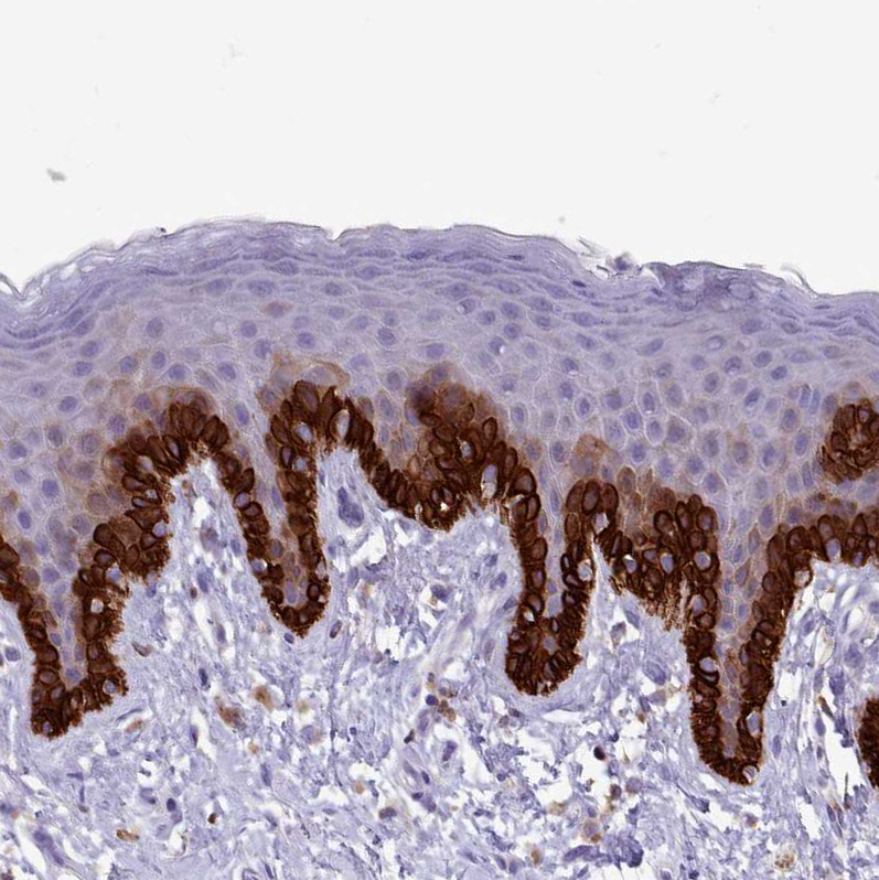
Immunohistochemical staining of human skin shows strong membranous positivity in squamous epithelial cells.

Immunohistochemical staining of human skin shows strong membranous positivity in squamous epithelial cells.
Anti-RAC1 Antibody
HPA047820
ApplicationsWestern Blot, ImmunoHistoChemistry
Product group Antibodies
ReactivityHuman
TargetRAC1
Overview
- SupplierAtlas Antibodies
- Product NameAnti-RAC1 Antibody
- Delivery Days Customer4
- ApplicationsWestern Blot, ImmunoHistoChemistry
- CertificationResearch Use Only
- ClonalityPolyclonal
- ConjugateUnconjugated
- Gene ID5879
- Target nameRAC1
- Target descriptionRac family small GTPase 1
- Target synonymsMIG5, MRD48, Rac-1, TC-25, p21-Rac1, ras-related C3 botulinum toxin substrate 1, cell migration-inducing gene 5 protein, ras-like protein TC25, ras-related C3 botulinum toxin substrate 1 (rho family, small GTP binding protein Rac1)
- HostRabbit
- IsotypeIgG
- Protein IDP63000
- Protein NameRas-related C3 botulinum toxin substrate 1
- Scientific DescriptionRecombinant Protein Epitope Signature Tag (PrEST) antigen sequence
- ReactivityHuman
- Storage Instruction-20°C,2°C to 8°C
- UNSPSC41116161
Datasheet
MSDS
Related products
Product group Antibodies
ApplicationsWestern Blot, ImmunoHistoChemistry
ReactivityHuman, Mouse, Rat
- SizePrice
Product group Antibodies
Anti-RAC1 Antibody130-10475
ApplicationsWestern Blot, ELISA
TargetRAC1
- SizePrice
Product group Antibodies
RAC1/2/3 Recombinant Antibody, AbBy Fluor-594 ConjugatedBSM-61458R-BF594
ApplicationsWestern Blot
ReactivityHuman, Mouse, Rat
TargetRAC1
- SizePrice
Product group Antibodies
Rac1 Polyclonal AntibodyCAC09043
ApplicationsImmunoFluorescence, ELISA, ImmunoHistoChemistry
TargetRAC1
- SizePrice
Product group Antibodies
RAC1 AntibodyCSB-PA04329A0RB
ApplicationsImmunoFluorescence, ELISA, ImmunoHistoChemistry
ReactivityHuman
TargetRAC1
- SizePrice
Product group Antibodies

RAC1 AntibodyLS-C409274
ApplicationsImmunoFluorescence, Western Blot, ImmunoHistoChemistry
ReactivityHuman, Mouse, Rat
TargetRAC1
- SizePrice
Product group Antibodies
Rac1 antibodyGTX100761
ApplicationsImmunoFluorescence, Western Blot, ImmunoCytoChemistry, ImmunoHistoChemistry, ImmunoHistoChemistry Paraffin
ReactivityHuman, Mouse, Rat
TargetRAC1
- SizePrice
Product group Antibodies
ApplicationsWestern Blot, ELISA
ReactivityHuman
TargetRAC1
- SizePrice