


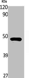
Figure 1. Western blot analysis of RASSF8 using anti-RASSF8 antibody (A11627-1). Electrophoresis was performed on a 5-20% SDS-PAGE gel at 70V (Stacking gel) / 90V (Resolving gel) for 2-3 hours. The sample well of each lane was loaded with 30 ug of sample under reducing conditions. Lane 1: human Hela whole cell lysates, Lane 2: human PC-3 whole cell lysates, Lane 3: human placenta tissue lysates, Lane 4: human U251 whole cell lysates, Lane 5: mouse testis tissue lysates, Lane 6: mouse lung tissue lysates. After electrophoresis, proteins were transferred to a nitrocellulose membrane at 150 mA for 50-90 minutes. Blocked the membrane with 5% non-fat milk/TBS for 1.5 hour at RT. The membrane was incubated with rabbit anti-RASSF8 antigen affinity purified polyclonal antibody (Catalog # A11627-1) at 0.5 microg/mL overnight at 4°C, then washed with TBS-0.1%Tween 3 times with 5 minutes each and probed with a goat anti-rabbit IgG-HRP secondary antibody at a dilution of 1:5000 for 1.5 hour at RT. The signal is developed using an Enhanced Chemiluminescent detection (ECL) kit (Catalog # EK1002) with Tanon 5200 system. A specific band was detected for RASSF8 at approximately 48 kDa. The expected band size for RASSF8 is at 48 kDa.
. RASSF8 was detected in an immunocytochemical section of HELA cells. Enzyme antigen retrieval was performed using IHC enzyme antigen retrieval reagent (AR0022) for 15 mins. The cells were blocked with 10% goat serum. And then incubated with 5 microg/mL rabbit anti-RASSF8 Antibody (A11627-1) overnight at 4°C. Cy3 Conjugated Goat Anti-Rabbit IgG (BA1032) was used as secondary antibody at 1:500 dilution and incubated for 30 minutes at 37°C. The section was counterstained with DAPI. Visualize using a fluorescence microscope and filter sets appropriate for the label used.")
. Overlay histogram showing PC-3 cells stained with A11627-1 (Blue line). To facilitate intracellular staining, cells were fixed with 4% paraformaldehyde and permeabilized with permeabilization buffer. The cells were blocked with 10% normal goat serum. And then incubated with rabbit anti-RASSF8 Antibody (A11627-1, 1 microg/1x106 cells) for 30 min at 20°C. DyLight®488 conjugated goat anti-rabbit IgG (BA1127, 5-10 microg/1x106 cells) was used as secondary antibody for 30 minutes at 20°C. Isotype control antibody (Green line) was rabbit IgG (1 microg/1x106) used under the same conditions. Unlabelled sample (Red line) was also used as a control.")
Figure 1. Western blot analysis of RASSF8 using anti-RASSF8 antibody (A11627-1). Electrophoresis was performed on a 5-20% SDS-PAGE gel at 70V (Stacking gel) / 90V (Resolving gel) for 2-3 hours. The sample well of each lane was loaded with 30 ug of sample under reducing conditions. Lane 1: human Hela whole cell lysates, Lane 2: human PC-3 whole cell lysates, Lane 3: human placenta tissue lysates, Lane 4: human U251 whole cell lysates, Lane 5: mouse testis tissue lysates, Lane 6: mouse lung tissue lysates. After electrophoresis, proteins were transferred to a nitrocellulose membrane at 150 mA for 50-90 minutes. Blocked the membrane with 5% non-fat milk/TBS for 1.5 hour at RT. The membrane was incubated with rabbit anti-RASSF8 antigen affinity purified polyclonal antibody (Catalog # A11627-1) at 0.5 microg/mL overnight at 4°C, then washed with TBS-0.1%Tween 3 times with 5 minutes each and probed with a goat anti-rabbit IgG-HRP secondary antibody at a dilution of 1:5000 for 1.5 hour at RT. The signal is developed using an Enhanced Chemiluminescent detection (ECL) kit (Catalog # EK1002) with Tanon 5200 system. A specific band was detected for RASSF8 at approximately 48 kDa. The expected band size for RASSF8 is at 48 kDa.
Anti-RASSF8 Antibody Picoband(r)
A11627-1-CARRIER-FREE
ApplicationsFlow Cytometry, ImmunoFluorescence, Western Blot, ELISA, ImmunoCytoChemistry
Product group Antibodies
ReactivityHuman, Mouse
TargetRASSF8
Overview
- SupplierBoster Bio
- Product NameAnti-RASSF8 Antibody Picoband(r)
- Delivery Days Customer9
- ApplicationsFlow Cytometry, ImmunoFluorescence, Western Blot, ELISA, ImmunoCytoChemistry
- CertificationResearch Use Only
- ClonalityPolyclonal
- Concentration500 ug/ml
- Gene ID11228
- Target nameRASSF8
- Target descriptionRas association domain family member 8
- Target synonymsC12orf2, HOJ1, ras association domain-containing protein 8, Ras association (RalGDS/AF-6) domain family (N-terminal) member 8, carcinoma-associated protein HOJ-1
- HostRabbit
- IsotypeIgG
- Protein IDQ8NHQ8
- Protein NameRas association domain-containing protein 8
- Scientific DescriptionBoster Bio Anti-RASSF8 Antibody Picoband® catalog # A11627-1. Tested in WB, ICC/IF, Flow Cytometry, ELISA applications. This antibody reacts with Human, Mouse. The brand Picoband indicates this is a premium antibody that guarantees superior quality, high affinity, and strong signals with minimal background in Western blot applications. Only our best-performing antibodies are designated as Picoband, ensuring unmatched performance.
- ReactivityHuman, Mouse
- Storage Instruction-20°C,2°C to 8°C
- UNSPSC12352203
Related products
Product group Antibodies
RASSF8 AntibodyCSB-PA004748
ApplicationsWestern Blot, ELISA
ReactivityHuman, Rat
TargetRASSF8
- SizePrice
Product group Antibodies
Anti-RASSF8 AntibodyA45741
ApplicationsImmunoHistoChemistry
ReactivityHuman
- SizePrice
Product group Antibodies

RASSF8 AntibodyLS-C831777
ApplicationsELISA, ImmunoHistoChemistry
ReactivityHuman, Mouse
TargetRASSF8
- SizePrice
Product group Antibodies

Goat anti-RASSF8EB08395
ApplicationsWestern Blot, ELISA
ReactivityBovine, Human, Mouse
TargetRASSF8
- SizePrice
Product group Antibodies
Anti-RASSF8 AntibodyHPA038163
ApplicationsImmunoHistoChemistry
ReactivityHuman
TargetRASSF8
- SizePrice
Product group Antibodies
RASSF8 antibodyGTX115947
ApplicationsWestern Blot
ReactivityHuman
TargetRASSF8
- SizePrice
Product group Antibodies
Anti-RASSF8 (N-term) Antibody102-20874
ApplicationsFlow Cytometry, Western Blot
TargetRASSF8
- SizePrice
Product group Antibodies
RASSF8 Polyclonal AntibodyBS-6089R
ApplicationsImmunoFluorescence, Western Blot, ELISA, ImmunoCytoChemistry, ImmunoHistoChemistry, ImmunoHistoChemistry Frozen, ImmunoHistoChemistry Paraffin
ReactivityBovine, Canine, Chicken, Equine, Human, Mouse, Porcine, Rat
TargetRASSF8
- SizePrice