


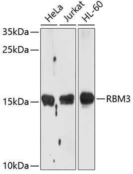
Figure 1. Western blot analysis of RBM3 using anti-RBM3 antibody (M03104). Electrophoresis was performed on a 5-20% SDS-PAGE gel at 70V (Stacking gel) / 90V (Resolving gel) for 2-3 hours. The sample well of each lane was loaded with 30 ug of sample under reducing conditions. Lane 1: human Jurkat whole cell lysates, Lane 2: human Hela whole cell lysates, Lane 3: human THP-1 whole cell lysates, Lane 4: human RT4 whole cell lysates. After electrophoresis, proteins were transferred to a nitrocellulose membrane at 150 mA for 50-90 minutes. Blocked the membrane with 5% non-fat milk/TBS for 1.5 hour at RT. The membrane was incubated with rabbit anti-RBM3 antigen affinity purified monoclonal antibody (Catalog # M03104) at 1:500 overnight at 4°C, then washed with TBS-0.1%Tween 3 times with 5 minutes each and probed with a goat anti-rabbit IgG-HRP secondary antibody at a dilution of 1:1000 for 1.5 hour at RT. The signal is developed using an Enhanced Chemiluminescent detection (ECL) kit (Catalog # EK1002) with Tanon 5200 system. A specific band was detected for RBM3 at approximately 17 kDa. The expected band size for RBM3 is at 17 kDa.
. RBM3 was detected in a paraffin-embedded section of human colorectal adenocarcinoma tissue. Heat mediated antigen retrieval was performed in EDTA buffer (pH 8.0, epitope retrieval solution). The tissue section was blocked with 10% goat serum. The tissue section was then incubated with 1:50 rabbit anti-RBM3 Antibody (M03104) overnight at 4°C. Peroxidase Conjugated Goat Anti-rabbit IgG was used as secondary antibody and incubated for 30 minutes at 37°C. The tissue section was developed using HRP Conjugated Rabbit IgG Super Vision Assay Kit (Catalog # SV0002) with DAB as the chromogen.")
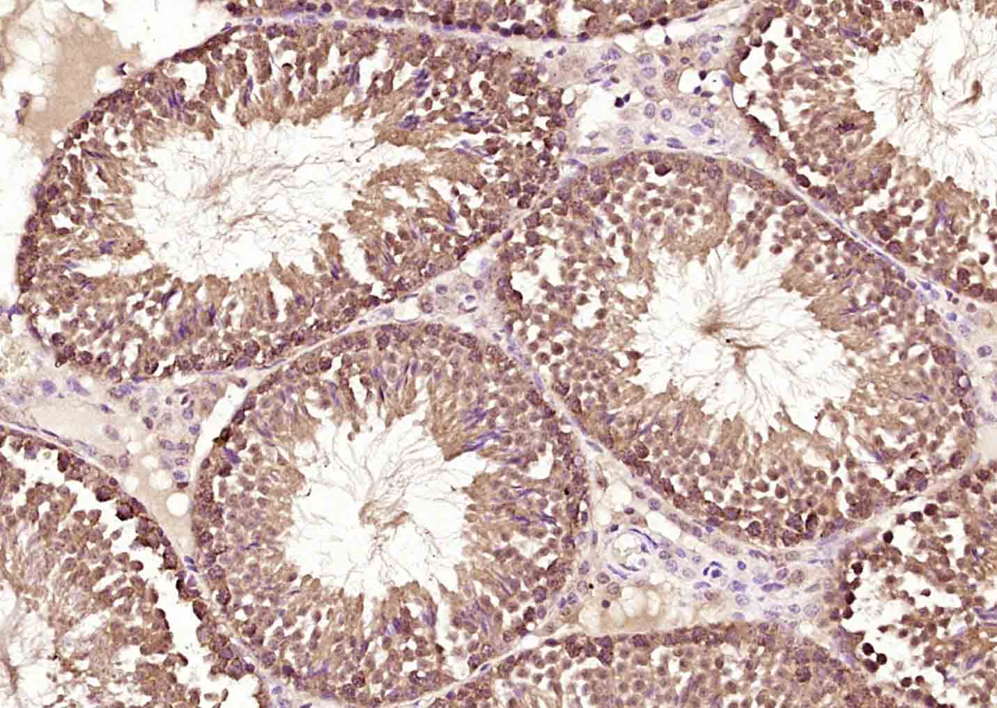
. RBM3 was detected in a paraffin-embedded section of human testicular germ cell tumor tissue. Heat mediated antigen retrieval was performed in EDTA buffer (pH 8.0, epitope retrieval solution). The tissue section was blocked with 10% goat serum. The tissue section was then incubated with 1:50 rabbit anti-RBM3 Antibody (M03104) overnight at 4°C. Peroxidase Conjugated Goat Anti-rabbit IgG was used as secondary antibody and incubated for 30 minutes at 37°C. The tissue section was developed using HRP Conjugated Rabbit IgG Super Vision Assay Kit (Catalog # SV0002) with DAB as the chromogen.")
. RBM3 was detected in a paraffin-embedded section of human liver cancer tissue. Heat mediated antigen retrieval was performed in EDTA buffer (pH 8.0, epitope retrieval solution). The tissue section was blocked with 10% goat serum. The tissue section was then incubated with 1:50 rabbit anti-RBM3 Antibody (M03104) overnight at 4°C. Peroxidase Conjugated Goat Anti-rabbit IgG was used as secondary antibody and incubated for 30 minutes at 37°C. The tissue section was developed using HRP Conjugated Rabbit IgG Super Vision Assay Kit (Catalog # SV0002) with DAB as the chromogen.")
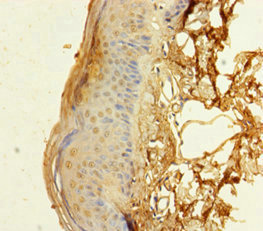
. RBM3 was detected in a paraffin-embedded section of human lung adenocarcinoma tissue. Heat mediated antigen retrieval was performed in EDTA buffer (pH 8.0, epitope retrieval solution). The tissue section was blocked with 10% goat serum. The tissue section was then incubated with 1:50 rabbit anti-RBM3 Antibody (M03104) overnight at 4°C. Peroxidase Conjugated Goat Anti-rabbit IgG was used as secondary antibody and incubated for 30 minutes at 37°C. The tissue section was developed using HRP Conjugated Rabbit IgG Super Vision Assay Kit (Catalog # SV0002) with DAB as the chromogen.")
. RBM3 was detected in a paraffin-embedded section of human prostate cancer tissue. Heat mediated antigen retrieval was performed in EDTA buffer (pH 8.0, epitope retrieval solution). The tissue section was blocked with 10% goat serum. The tissue section was then incubated with 1:50 rabbit anti-RBM3 Antibody (M03104) overnight at 4°C. Peroxidase Conjugated Goat Anti-rabbit IgG was used as secondary antibody and incubated for 30 minutes at 37°C. The tissue section was developed using HRP Conjugated Rabbit IgG Super Vision Assay Kit (Catalog # SV0002) with DAB as the chromogen.")
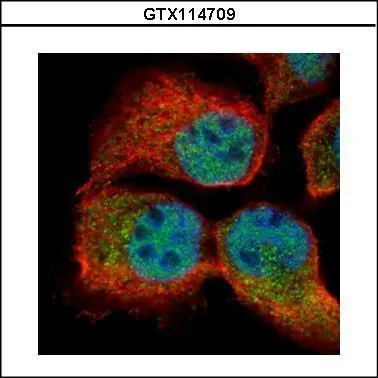
 and anti-Beta Tubulin antibody (M01857-3). RBM3 was detected in immunocytochemical section of A549 cell. Enzyme antigen retrieval was performed using IHC enzyme antigen retrieval reagent (AR0022) for 15 mins. The cells were blocked with 10% goat serum. And then incubated at 1:50 with rabbit anti-RBM3 Antibody (M03104) and mouse anti-Beta Tubulin antibody (M01857-3) overnight at 4°C. DyLight?488 Conjugated Goat Anti-Rabbit IgG (BA1127) and Cy3 Conjugated Goat Anti-Mouse IgG (BA1031) were used as secondary antibody at 1:500 dilution and incubated for 30 minutes at 37°C. Visualize using a fluorescence microscope and filter sets appropriate for the label used.")
Figure 1. Western blot analysis of RBM3 using anti-RBM3 antibody (M03104). Electrophoresis was performed on a 5-20% SDS-PAGE gel at 70V (Stacking gel) / 90V (Resolving gel) for 2-3 hours. The sample well of each lane was loaded with 30 ug of sample under reducing conditions. Lane 1: human Jurkat whole cell lysates, Lane 2: human Hela whole cell lysates, Lane 3: human THP-1 whole cell lysates, Lane 4: human RT4 whole cell lysates. After electrophoresis, proteins were transferred to a nitrocellulose membrane at 150 mA for 50-90 minutes. Blocked the membrane with 5% non-fat milk/TBS for 1.5 hour at RT. The membrane was incubated with rabbit anti-RBM3 antigen affinity purified monoclonal antibody (Catalog # M03104) at 1:500 overnight at 4°C, then washed with TBS-0.1%Tween 3 times with 5 minutes each and probed with a goat anti-rabbit IgG-HRP secondary antibody at a dilution of 1:1000 for 1.5 hour at RT. The signal is developed using an Enhanced Chemiluminescent detection (ECL) kit (Catalog # EK1002) with Tanon 5200 system. A specific band was detected for RBM3 at approximately 17 kDa. The expected band size for RBM3 is at 17 kDa.
Anti-RBM3 Rabbit Monoclonal Antibody
M03104
ApplicationsFlow Cytometry, ImmunoFluorescence, Western Blot, ImmunoCytoChemistry, ImmunoHistoChemistry
Product group Antibodies
ReactivityHuman, Mouse
TargetRBM3
Overview
- SupplierBoster Bio
- Product NameAnti-RBM3 Rabbit Monoclonal Antibody
- Delivery Days Customer9
- ApplicationsFlow Cytometry, ImmunoFluorescence, Western Blot, ImmunoCytoChemistry, ImmunoHistoChemistry
- CertificationResearch Use Only
- ClonalityMonoclonal
- Clone ID29R08
- Gene ID5935
- Target nameRBM3
- Target descriptionRNA binding motif protein 3
- Target synonymsIS1-RNPL, RNPL, RNA-binding protein 3, RNA binding motif (RNP1, RRM) protein 3
- HostRabbit
- IsotypeIgG
- Protein IDP98179
- Protein NameRNA-binding protein 3
- Scientific DescriptionBoster Bio Anti-RBM3 Rabbit Monoclonal Antibody catalog # M03104. Tested in WB, IHC, ICC/IF, Flow Cytometry applications. This antibody reacts with Human, Mouse.
- ReactivityHuman, Mouse
- Storage Instruction-20°C
- UNSPSC12352203
Related products
Product group Antibodies
Anti-RBM3 AntibodyA11899
ApplicationsImmunoFluorescence, Western Blot, ImmunoCytoChemistry, ImmunoHistoChemistry
ReactivityHuman, Mouse, Rat
- SizePrice
Product group Antibodies
Anti-RBM3 Antibody144-05931
ApplicationsWestern Blot, ImmunoHistoChemistry
ReactivityHuman, Mouse, Rat
TargetRBM3
- SizePrice

Product group Antibodies
Anti-RBM3 AntibodyAMAB90655
ApplicationsWestern Blot, ImmunoCytoChemistry, ImmunoHistoChemistry
ReactivityHuman
TargetRBM3
- SizePrice
Product group Antibodies

RBM3 AntibodyLS-C831472
ApplicationsImmunoHistoChemistry
ReactivityHuman
TargetRBM3
- SizePrice
Product group Antibodies
RBM3 Polyclonal AntibodyBS-5902R
ApplicationsImmunoFluorescence, Western Blot, ELISA, ImmunoCytoChemistry, ImmunoHistoChemistry, ImmunoHistoChemistry Frozen, ImmunoHistoChemistry Paraffin
ReactivityCanine, Equine, Human, Mouse, Porcine, Rabbit, Rat
TargetRBM3
- SizePrice
Product group Antibodies

Goat anti-RBM3EB09201
ApplicationsWestern Blot, ELISA
ReactivityCanine, Human, Mouse, Rat
TargetRBM3
- SizePrice
Product group Antibodies
RBM3 Polyclonal AntibodyCAC13181
ApplicationsELISA, ImmunoHistoChemistry
TargetRBM3
- SizePrice
Product group Antibodies
RBM3 AntibodyCSB-PA019420HA01HU
ApplicationsELISA, ImmunoHistoChemistry
ReactivityHuman
TargetRBM3
- SizePrice
Product group Antibodies
RBM3 antibodyGTX114709
ApplicationsImmunoFluorescence, ImmunoCytoChemistry
ReactivityHuman
TargetRBM3
- SizePrice
Product group Antibodies
Anti-RBM3 AntibodyCAB5931
ApplicationsWestern Blot, ELISA, ImmunoHistoChemistry, ImmunoHistoChemistry Paraffin
ReactivityHuman
TargetRBM3
- SizePrice