


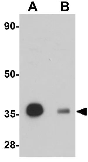
Figure 1. Western blot analysis of REEP3 using anti-REEP3 antibody (A12243-1). Electrophoresis was performed on a 5-20% SDS-PAGE gel at 70V (Stacking gel) / 90V (Resolving gel) for 2-3 hours. The sample well of each lane was loaded with 30 ug of sample under reducing conditions. Lane 1: human Hacat whole cell lysates, Lane 2: human PC-3 whole cell lysates, Lane 3: human A431 whole cell lysates, Lane 4: human SIHA whole cell lysates, Lane 5: rat brain tissue lysates, Lane 6: rat PC-12 whole cell lysates, Lane 7: mouse brain tissue lysates, Lane 8: mouse NIH/3T3 whole cell lysates. After electrophoresis, proteins were transferred to a nitrocellulose membrane at 150 mA for 50-90 minutes. Blocked the membrane with 5% non-fat milk/TBS for 1.5 hour at RT. The membrane was incubated with rabbit anti-REEP3 antigen affinity purified polyclonal antibody (Catalog # A12243-1) at 0.5 microg/mL overnight at 4°C, then washed with TBS-0.1%Tween 3 times with 5 minutes each and probed with a goat anti-rabbit IgG-HRP secondary antibody at a dilution of 1:5000 for 1.5 hour at RT. The signal is developed using an Enhanced Chemiluminescent detection (ECL) kit (Catalog # EK1002) with Tanon 5200 system. A specific band was detected for REEP3 at approximately 34 kDa. The expected band size for REEP3 is at 29 kDa.
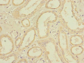
. REEP3 was detected in an immunocytochemical section of U2OS cells. Enzyme antigen retrieval was performed using IHC enzyme antigen retrieval reagent (AR0022) for 15 mins. The cells were blocked with 10% goat serum. And then incubated with 5 microg/mL rabbit anti-REEP3 Antibody (A12243-1) overnight at 4°C. Cy3 Conjugated Goat Anti-Rabbit IgG (BA1032) was used as secondary antibody at 1:500 dilution and incubated for 30 minutes at 37°C. The section was counterstained with DAPI. Visualize using a fluorescence microscope and filter sets appropriate for the label used.")
Figure 1. Western blot analysis of REEP3 using anti-REEP3 antibody (A12243-1). Electrophoresis was performed on a 5-20% SDS-PAGE gel at 70V (Stacking gel) / 90V (Resolving gel) for 2-3 hours. The sample well of each lane was loaded with 30 ug of sample under reducing conditions. Lane 1: human Hacat whole cell lysates, Lane 2: human PC-3 whole cell lysates, Lane 3: human A431 whole cell lysates, Lane 4: human SIHA whole cell lysates, Lane 5: rat brain tissue lysates, Lane 6: rat PC-12 whole cell lysates, Lane 7: mouse brain tissue lysates, Lane 8: mouse NIH/3T3 whole cell lysates. After electrophoresis, proteins were transferred to a nitrocellulose membrane at 150 mA for 50-90 minutes. Blocked the membrane with 5% non-fat milk/TBS for 1.5 hour at RT. The membrane was incubated with rabbit anti-REEP3 antigen affinity purified polyclonal antibody (Catalog # A12243-1) at 0.5 microg/mL overnight at 4°C, then washed with TBS-0.1%Tween 3 times with 5 minutes each and probed with a goat anti-rabbit IgG-HRP secondary antibody at a dilution of 1:5000 for 1.5 hour at RT. The signal is developed using an Enhanced Chemiluminescent detection (ECL) kit (Catalog # EK1002) with Tanon 5200 system. A specific band was detected for REEP3 at approximately 34 kDa. The expected band size for REEP3 is at 29 kDa.
Anti-REEP3 Antibody Picoband(r)
A12243-1-CARRIER-FREE
ApplicationsImmunoFluorescence, Western Blot, ImmunoCytoChemistry
Product group Antibodies
ReactivityHuman, Mouse, Rat
TargetREEP3
Overview
- SupplierBoster Bio
- Product NameAnti-REEP3 Antibody Picoband(r)
- Delivery Days Customer9
- ApplicationsImmunoFluorescence, Western Blot, ImmunoCytoChemistry
- CertificationResearch Use Only
- ClonalityPolyclonal
- Concentration500 ug/ml
- Gene ID221035
- Target nameREEP3
- Target descriptionreceptor accessory protein 3
- Target synonymsC10orf74, Yip2b, receptor expression-enhancing protein 3
- HostRabbit
- IsotypeIgG
- Protein IDQ6NUK4
- Protein NameReceptor expression-enhancing protein 3
- Scientific DescriptionBoster Bio Anti-REEP3 Antibody Picoband® catalog # A12243-1. Tested in IF, ICC, WB applications. This antibody reacts with Human, Mouse, Rat. The brand Picoband indicates this is a premium antibody that guarantees superior quality, high affinity, and strong signals with minimal background in Western blot applications. Only our best-performing antibodies are designated as Picoband, ensuring unmatched performance.
- ReactivityHuman, Mouse, Rat
- Storage Instruction-20°C,2°C to 8°C
- UNSPSC12352203
Related products
Product group Antibodies
Anti-REEP3 AntibodyA48025
ApplicationsWestern Blot, ELISA, ImmunoHistoChemistry
ReactivityHuman, Mouse, Rat
- SizePrice

Product group Antibodies
REEP3 AntibodyCSB-PA754137LA01HU
ApplicationsImmunoFluorescence, ELISA, ImmunoHistoChemistry
ReactivityHuman
TargetREEP3
- SizePrice
Product group Antibodies
REEP3 antibodyGTX85210
ApplicationsImmunoFluorescence, Western Blot, ELISA, ImmunoCytoChemistry, ImmunoHistoChemistry, ImmunoHistoChemistry Paraffin
ReactivityHuman, Mouse, Rat
TargetREEP3
- SizePrice
Product group Antibodies

REEP3 Antibody (Biotin)LS-C399265
ApplicationsELISA
ReactivityHuman
TargetREEP3
- SizePrice
Product group Antibodies
REEP3 AntibodyPACO36962
ApplicationsImmunoFluorescence, ELISA, ImmunoHistoChemistry
ReactivityHuman
TargetREEP3
- SizePrice