


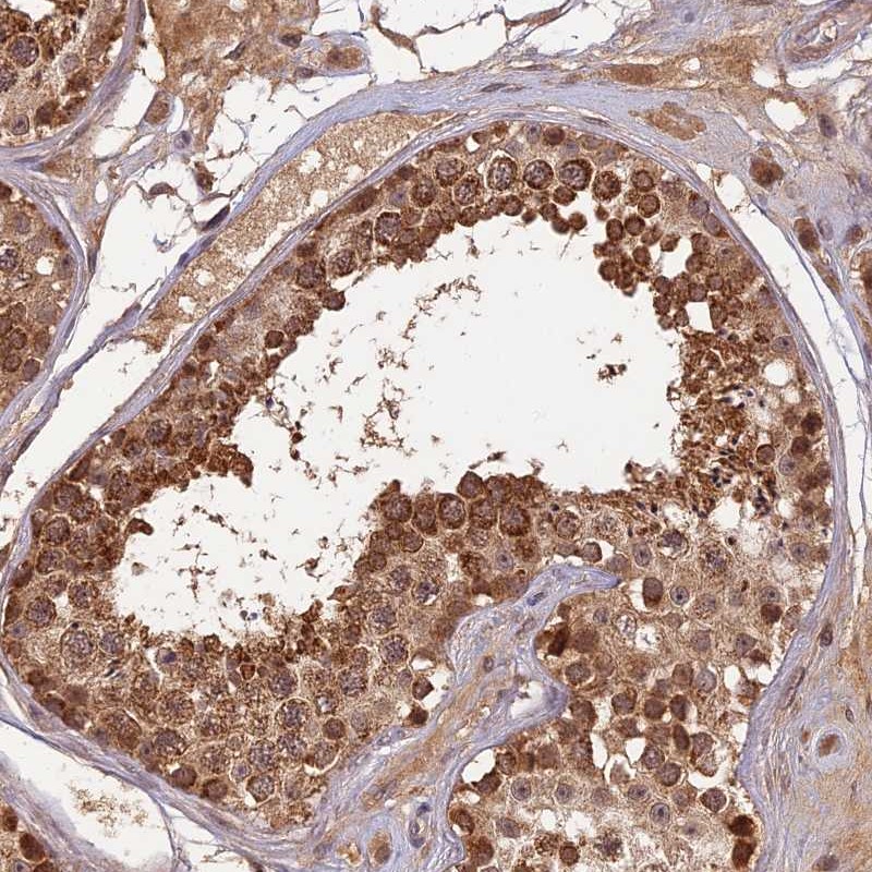
Immunohistochemical staining of human kidney shows strong positivity in cells in glomeruli and moderate staining in cells in tubules.
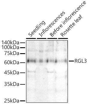
 and RGL3 over-expression lysate (Co-expressed with a C-terminal myc-DDK tag (~3.1 kDa) in mammalian HEK293T cells, LY422124).")
Immunohistochemical staining of human kidney shows strong positivity in cells in glomeruli and moderate staining in cells in tubules.
Anti-RGL3 Antibody
HPA042763
ApplicationsWestern Blot, ImmunoHistoChemistry
Product group Antibodies
ReactivityHuman
TargetRGL3
Overview
- SupplierAtlas Antibodies
- Product NameAnti-RGL3 Antibody
- Delivery Days Customer4
- ApplicationsWestern Blot, ImmunoHistoChemistry
- CertificationResearch Use Only
- ClonalityPolyclonal
- ConjugateUnconjugated
- Gene ID57139
- Target nameRGL3
- Target descriptionral guanine nucleotide dissociation stimulator like 3
- Target synonymsral guanine nucleotide dissociation stimulator-like 3, RalGEF-like protein 3, mouse homolog, ralGDS-like 3
- HostRabbit
- IsotypeIgG
- Protein IDQ3MIN7
- Protein NameRal guanine nucleotide dissociation stimulator-like 3
- Scientific DescriptionRecombinant Protein Epitope Signature Tag (PrEST) antigen sequence
- ReactivityHuman
- Storage Instruction-20°C,2°C to 8°C
- UNSPSC41116161
Datasheet
MSDS
Related products

Product group Antibodies
Anti-RGL3 Antibody Picoband(r)A11774-1-CARRIER-FREE
ApplicationsFlow Cytometry, ImmunoFluorescence, Western Blot, ELISA, ImmunoCytoChemistry, ImmunoHistoChemistry
ReactivityHuman
TargetRGL3
- SizePrice
Product group Antibodies
Anti-RGL3 AntibodyHPA043615
ApplicationsWestern Blot, ImmunoHistoChemistry
ReactivityHuman
TargetRGL3
- SizePrice
Product group Antibodies
Anti-RGL3 AntibodyHPA043615
ApplicationsWestern Blot, ImmunoHistoChemistry
ReactivityHuman
TargetRGL3
- SizePrice
Product group Antibodies

RGL3 Antibody (aa338-387)LS-C135374
ApplicationsWestern Blot
ReactivityHuman
TargetRGL3
- SizePrice