

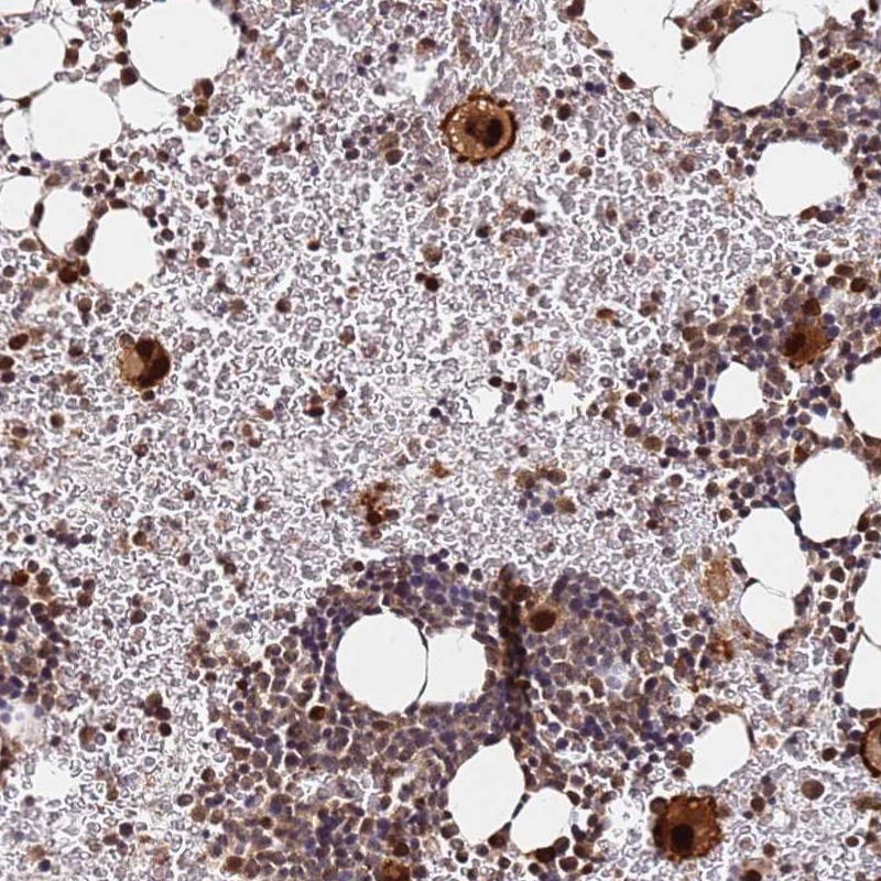
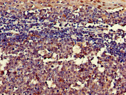
Immunohistochemical staining of human bone marrow shows strong nuclear positivity in megakaryocytes.
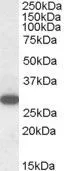
 and RGS18 over-expression lysate (Co-expressed with a C-terminal myc-DDK tag (~3.1 kDa) in mammalian HEK293T cells, LY408941).")
Immunohistochemical staining of human bone marrow shows strong nuclear positivity in megakaryocytes.
Anti-RGS18 Antibody
HPA045780
ApplicationsWestern Blot, ImmunoHistoChemistry
Product group Antibodies
ReactivityHuman
TargetRGS18
Overview
- SupplierAtlas Antibodies
- Product NameAnti-RGS18 Antibody
- Delivery Days Customer4
- ApplicationsWestern Blot, ImmunoHistoChemistry
- CertificationResearch Use Only
- ClonalityPolyclonal
- ConjugateUnconjugated
- Gene ID64407
- Target nameRGS18
- Target descriptionregulator of G protein signaling 18
- Target synonymsRGS13, regulator of G-protein signaling 18, regulator of G-protein signalling 13, regulator of G-protein signalling 18
- HostRabbit
- IsotypeIgG
- Protein IDQ9NS28
- Protein NameRegulator of G-protein signaling 18
- Scientific DescriptionRecombinant Protein Epitope Signature Tag (PrEST) antigen sequence
- ReactivityHuman
- Storage Instruction-20°C,2°C to 8°C
- UNSPSC41116161
Datasheet
MSDS
Related products

Product group Antibodies
Anti-RGS18 AntibodyA84027
ApplicationsWestern Blot, ELISA, ImmunoHistoChemistry
ReactivityHuman
- SizePrice

Product group Antibodies
Anti-RGS18 Antibody Picoband(r)A10844-2-CARRIER-FREE
ApplicationsFlow Cytometry, Western Blot, ELISA
ReactivityHuman
TargetRGS18
- SizePrice
Product group Antibodies
RGS18 Recombinant AntibodyBSM-62108R
ApplicationsWestern Blot
ReactivityHuman
TargetRGS18
- SizePrice
Product group Antibodies

Goat anti-RGS18EB06372
ApplicationsWestern Blot, ELISA, ImmunoHistoChemistry
ReactivityHuman
TargetRGS18
- SizePrice
Product group Antibodies
RGS18 AntibodyCSB-PA019649LA01HU
ApplicationsImmunoFluorescence, ELISA, ImmunoHistoChemistry
ReactivityHuman
TargetRGS18
- SizePrice
Product group Antibodies

RGS18 Antibody (Biotin)LS-C499658
ApplicationsELISA
ReactivityHuman
TargetRGS18
- SizePrice
Product group Antibodies
RGS18 antibody, C-termGTX89731
ApplicationsWestern Blot
ReactivityHuman
TargetRGS18
- SizePrice

Product group Antibodies
Anti-RGS18 AntibodyHPA028727
ApplicationsImmunoCytoChemistry
ReactivityHuman
TargetRGS18
- SizePrice