

Anti-RHOT1 (N-term) Antibody
102-23243
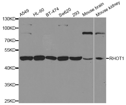
ApplicationsWestern Blot
Product group Antibodies
TargetRHOT1
Overview
- SupplierRayBiotech
- Product NameAnti-RHOT1 (N-term) Antibody
- Delivery Days Customer16
- ApplicationsWestern Blot
- CertificationResearch Use Only
- ClonalityPolyclonal
- Clone IDRB38600
- Gene ID55288
- Target nameRHOT1
- Target descriptionras homolog family member T1
- Target synonymsARHT1, MIRO-1, MIRO1, mitochondrial Rho GTPase 1, mitochondrial Rho (MIRO) GTPase 1, rac-GTP binding protein-like protein, ras homolog gene family, member T1
- HostRabbit
- Protein IDQ2HJF8
- Protein NameMitochondrial Rho GTPase 1
- Scientific DescriptionRabbit Anti-RHOT1 (N-term) Antibody, 400 microl
- Storage Instruction-20°C
- UNSPSC12352203
Related products
Product group Antibodies
Anti-RHOT1 AntibodyA31083
ApplicationsWestern Blot, ImmunoHistoChemistry
ReactivityHuman, Mouse, Rat
- SizePrice

Product group Antibodies
Anti-RHOT1 AntibodyAMAB90854
ApplicationsImmunoHistoChemistry
ReactivityHuman
TargetRHOT1
- SizePrice

Product group Antibodies
RHOT1 antibodyGTX31938
ApplicationsWestern Blot, ELISA, ImmunoHistoChemistry, ImmunoHistoChemistry Paraffin
ReactivityHuman, Mouse, Rat
TargetRHOT1
- SizePrice
Product group Antibodies

RHOT1 AntibodyLS-C334329
ApplicationsWestern Blot, ImmunoHistoChemistry
ReactivityHuman, Mouse, Rat
TargetRHOT1
- SizePrice

Product group Antibodies
Anti-MIRO1/RHOT1 Antibody Picoband(r)A05928-1-CARRIER-FREE
ApplicationsImmunoFluorescence, Western Blot, ELISA, ImmunoCytoChemistry, ImmunoHistoChemistry
ReactivityHuman, Mouse, Rat
TargetRHOT1
- SizePrice