

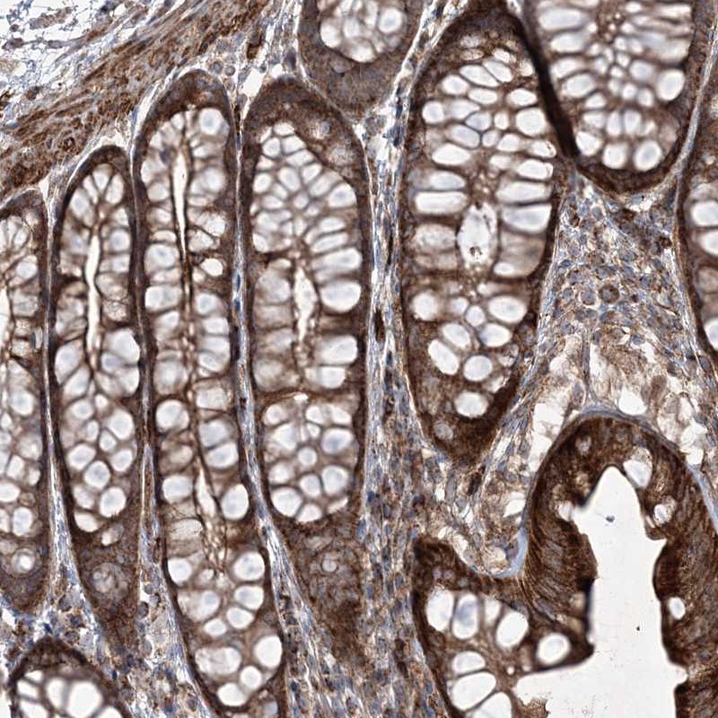
Immunohistochemical staining of human colon shows strong cytoplasmic and membranous positivity in glandular cells.
Immunohistochemical staining of human colon shows strong cytoplasmic and membranous positivity in glandular cells.
Anti-RICTOR Antibody
HPA037803
ApplicationsImmunoHistoChemistry
Product group Antibodies
ReactivityHuman
TargetRICTOR
Overview
- SupplierAtlas Antibodies
- Product NameAnti-RICTOR Antibody
- Delivery Days Customer4
- ApplicationsImmunoHistoChemistry
- CertificationResearch Use Only
- ClonalityPolyclonal
- ConjugateUnconjugated
- Gene ID253260
- Target nameRICTOR
- Target descriptionRPTOR independent companion of MTOR complex 2
- Target synonymsAVO3, PIA, hAVO3, rapamycin-insensitive companion of mTOR, AVO3 homolog, TORC2-specific protein AVO3, pianissimo
- HostRabbit
- IsotypeIgG
- Protein IDQ6R327
- Protein NameRapamycin-insensitive companion of mTOR
- Scientific DescriptionRecombinant Protein Epitope Signature Tag (PrEST) antigen sequence
- ReactivityHuman
- Storage Instruction-20°C,2°C to 8°C
- UNSPSC41116161
Datasheet
MSDS
Related products

Product group Antibodies
Anti-RICTOR Antibody Picoband(r)A03195-1-CARRIER-FREE
ApplicationsFlow Cytometry, ImmunoFluorescence, Western Blot, ELISA, ImmunoCytoChemistry, ImmunoHistoChemistry
ReactivityHuman
TargetRICTOR
- SizePrice
Product group Antibodies
Anti-RICTOR Antibody144-66055
ApplicationsWestern Blot
ReactivityHuman, Mouse, Rat
TargetRICTOR
- SizePrice

Product group Antibodies
RICTOR AntibodyCSB-PA108659
ApplicationsELISA, ImmunoHistoChemistry
ReactivityHuman, Mouse
TargetRICTOR
- SizePrice
Product group Antibodies

RICTOR AntibodyLS-C401351
ApplicationsELISA, ImmunoHistoChemistry
ReactivityHuman, Mouse
TargetRICTOR
- SizePrice
![WB analysis of HeLa (1), PANC-1 (2), MOLT4 (3), and HepG2 (4) cell lysate using GTX82786 RICTOR antibody [4H5].](https://www.genetex.com/upload/website/prouct_img/normal/GTX82786/GTX82786_20170912_WB_w_23061322_116.webp)
Product group Antibodies
RICTOR antibody [4H5]GTX82786
ApplicationsWestern Blot, ELISA
ReactivityHuman, Monkey, Mouse
TargetRICTOR
- SizePrice