


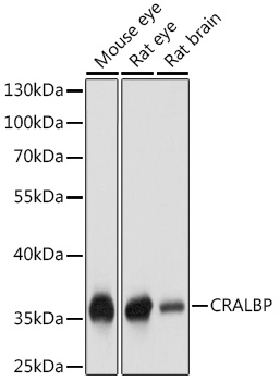
Figure 1. Western blot analysis of RLBP1 using anti-RLBP1 antibody (A05421-1). Electrophoresis was performed on a 5-20% SDS-PAGE gel at 70V (Stacking gel) / 90V (Resolving gel) for 2-3 hours. The sample well of each lane was loaded with 30 ug of sample under reducing conditions. Lane 1: rat eye tissue lysates, Lane 2: mouse eye tissue lysates. After electrophoresis, proteins were transferred to a nitrocellulose membrane at 150 mA for 50-90 minutes. Blocked the membrane with 5% non-fat milk/TBS for 1.5 hour at RT. The membrane was incubated with rabbit anti-RLBP1 antigen affinity purified polyclonal antibody (Catalog # A05421-1) at 0.5 microg/mL overnight at 4°C, then washed with TBS-0.1%Tween 3 times with 5 minutes each and probed with a goat anti-rabbit IgG-HRP secondary antibody at a dilution of 1:5000 for 1.5 hour at RT. The signal is developed using an Enhanced Chemiluminescent detection (ECL) kit (Catalog # EK1002) with Tanon 5200 system. A specific band was detected for RLBP1 at approximately 36 kDa. The expected band size for RLBP1 is at 36 kDa.
. Overlay histogram showing HepG2 cells stained with A05421-1 (Blue line). To facilitate intracellular staining, cells were fixed with 4% paraformaldehyde and permeabilized with permeabilization buffer. The cells were blocked with 10% normal goat serum. And then incubated with rabbit anti-RLBP1 Antibody (A05421-1, 1 microg/1x106 cells) for 30 min at 20°C. DyLight®488 conjugated goat anti-rabbit IgG (BA1127, 5-10 microg/1x106 cells) was used as secondary antibody for 30 minutes at 20°C. Isotype control antibody (Green line) was rabbit IgG (1 microg/1x106) used under the same conditions. Unlabelled sample without incubation with primary antibody and secondary antibody (Red line) was used as a blank control.")
Figure 1. Western blot analysis of RLBP1 using anti-RLBP1 antibody (A05421-1). Electrophoresis was performed on a 5-20% SDS-PAGE gel at 70V (Stacking gel) / 90V (Resolving gel) for 2-3 hours. The sample well of each lane was loaded with 30 ug of sample under reducing conditions. Lane 1: rat eye tissue lysates, Lane 2: mouse eye tissue lysates. After electrophoresis, proteins were transferred to a nitrocellulose membrane at 150 mA for 50-90 minutes. Blocked the membrane with 5% non-fat milk/TBS for 1.5 hour at RT. The membrane was incubated with rabbit anti-RLBP1 antigen affinity purified polyclonal antibody (Catalog # A05421-1) at 0.5 microg/mL overnight at 4°C, then washed with TBS-0.1%Tween 3 times with 5 minutes each and probed with a goat anti-rabbit IgG-HRP secondary antibody at a dilution of 1:5000 for 1.5 hour at RT. The signal is developed using an Enhanced Chemiluminescent detection (ECL) kit (Catalog # EK1002) with Tanon 5200 system. A specific band was detected for RLBP1 at approximately 36 kDa. The expected band size for RLBP1 is at 36 kDa.
Anti-RLBP1 Antibody Picoband(r)
A05421-1-CARRIER-FREE
ApplicationsFlow Cytometry, Western Blot, ELISA
Product group Antibodies
ReactivityHuman, Mouse, Rat
TargetRLBP1
Overview
- SupplierBoster Bio
- Product NameAnti-RLBP1 Antibody Picoband(r)
- Delivery Days Customer9
- ApplicationsFlow Cytometry, Western Blot, ELISA
- CertificationResearch Use Only
- ClonalityPolyclonal
- Concentration500 ug/ml
- Gene ID6017
- Target nameRLBP1
- Target descriptionretinaldehyde binding protein 1
- Target synonymsCRALBP, retinaldehyde-binding protein 1, cellular retinaldehyde-binding protein, cellular retinaldehyde-binding protein-1
- HostRabbit
- IsotypeIgG
- Protein IDP12271
- Protein NameRetinaldehyde-binding protein 1
- Scientific DescriptionBoster Bio Anti-RLBP1 Antibody Picoband® catalog # A05421-1. Tested in ELISA, Flow Cytometry, WB applications. This antibody reacts with Human, Mouse, Rat. The brand Picoband indicates this is a premium antibody that guarantees superior quality, high affinity, and strong signals with minimal background in Western blot applications. Only our best-performing antibodies are designated as Picoband, ensuring unmatched performance.
- ReactivityHuman, Mouse, Rat
- Storage Instruction-20°C,2°C to 8°C
- UNSPSC12352203
Related products
Product group Antibodies
Anti-CRALBP AntibodyA17253
ApplicationsWestern Blot
ReactivityMouse, Rat
- SizePrice
Product group Antibodies
Anti-Mouse/Rat RLBP1 Antibody144-09094
ApplicationsWestern Blot
ReactivityHuman, Mouse, Rat
TargetRLBP1
- SizePrice
Product group Antibodies
CRALBP Recombinant AntibodyBSM-60602R
ApplicationsImmunoFluorescence, Western Blot, ImmunoHistoChemistry, ImmunoHistoChemistry Frozen, ImmunoHistoChemistry Paraffin
ReactivityMouse, Rat
TargetRLBP1
- SizePrice
Product group Antibodies
ApplicationsImmunoPrecipitation, Western Blot, ImmunoCytoChemistry, ImmunoHistoChemistry
ReactivityMouse, Porcine, Rat
TargetRLBP1
- SizePrice
Product group Antibodies
RLBP1 AntibodyCSB-PA019743LA01HU
ApplicationsImmunoFluorescence, ELISA, ImmunoHistoChemistry
ReactivityHuman
TargetRLBP1
- SizePrice
Product group Antibodies

RLBP1 / CRALBP AntibodyLS-C410628
ApplicationsWestern Blot, ImmunoHistoChemistry, ImmunoHistoChemistry Paraffin
ReactivityHuman, Mouse, Rat
TargetRLBP1
- SizePrice

Product group Antibodies
Anti-RLBP1 AntibodyHPA044083
ApplicationsImmunoCytoChemistry, ImmunoHistoChemistry
ReactivityHuman
TargetRLBP1
- SizePrice
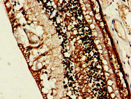
![CRALBP antibody [N2C3] detects CRALBP protein by immunohistochemical analysis. Sample: Paraffin-embedded rat eye. Green: CRALBP stained by CRALBP antibody [N2C3] (GTX113541) diluted at 1:500. Red: beta Tubulin 3/ Tuj1 , a Cytoskeleton marker, stained by beta Tubulin 3/ Tuj1 antibody [GT11710] (GTX631836) diluted at 1:500. Blue: Fluoroshield with DAPI (GTX30920). Antigen Retrieval: Citrate buffer, pH 6.0, 15 min](https://www.genetex.com/upload/website/prouct_img/normal/GTX113541/GTX113541_44510_20230505_IHC-P_R_23050918_270.webp)
Product group Antibodies
CRALBP antibody [N2C3]GTX113541
ApplicationsImmunoFluorescence, Western Blot, ImmunoCytoChemistry, ImmunoHistoChemistry, ImmunoHistoChemistry Paraffin
ReactivityHuman, Mouse, Rat
TargetRLBP1
- SizePrice