


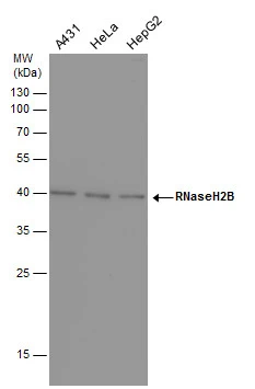
Figure 1. Western blot analysis of RNASEH2B using anti-RNASEH2B antibody (A06660-1). Electrophoresis was performed on a 5-20% SDS-PAGE gel at 70V (Stacking gel) / 90V (Resolving gel) for 2-3 hours. The sample well of each lane was loaded with 30 ug of sample under reducing conditions. Lane 1: human MOLT-4 whole cell lysates. After electrophoresis, proteins were transferred to a nitrocellulose membrane at 150 mA for 50-90 minutes. Blocked the membrane with 5% non-fat milk/TBS for 1.5 hour at RT. The membrane was incubated with rabbit anti-RNASEH2B antigen affinity purified polyclonal antibody (Catalog # A06660-1) at 0.5 microg/mL overnight at 4°C, then washed with TBS-0.1%Tween 3 times with 5 minutes each and probed with a goat anti-rabbit IgG-HRP secondary antibody at a dilution of 1:5000 for 1.5 hour at RT. The signal is developed using an Enhanced Chemiluminescent detection (ECL) kit (Catalog # EK1002) with Tanon 5200 system. A specific band was detected for RNASEH2B at approximately 35 kDa. The expected band size for RNASEH2B is at 35 kDa.
 and anti-Tubulin Alpha antibody (M03989-3). RNASEH2B was detected in immunocytochemical section of T-47D cell. Enzyme antigen retrieval was performed using IHC enzyme antigen retrieval reagent (AR0022) for 15 mins. The cells were blocked with 10% goat serum. And then incubated with 5 microg/mL rabbit anti-RNASEH2B Antibody (A06660-1) and mouse anti-Tubulin Alpha antibody (M03989-3) overnight at 4°C. Cy3 Conjugated Goat Anti-Rabbit IgG (BA1032) and DyLight®488 Conjugated Goat Anti-Mouse IgG (BA1126) were used as secondary antibody at 1:500 dilution and incubated for 30 minutes at 37°C. Visualize using a fluorescence microscope and filter sets appropriate for the label used.")
. Overlay histogram showing HL-60 cells stained with A06660-1 (Blue line). To facilitate intracellular staining, cells were fixed with 4% paraformaldehyde and permeabilized with permeabilization buffer. The cells were blocked with 10% normal goat serum. And then incubated with rabbit anti-RNASEH2B Antibody (A06660-1, 1 microg/1x106 cells) for 30 min at 20°C. DyLight®488 conjugated goat anti-rabbit IgG (BA1127, 5-10 microg/1x106 cells) was used as secondary antibody for 30 minutes at 20°C. Isotype control antibody (Green line) was rabbit IgG (1 microg/1x106) used under the same conditions. Unlabelled sample without incubation with primary antibody and secondary antibody (Red line) was used as a blank control.")
Figure 1. Western blot analysis of RNASEH2B using anti-RNASEH2B antibody (A06660-1). Electrophoresis was performed on a 5-20% SDS-PAGE gel at 70V (Stacking gel) / 90V (Resolving gel) for 2-3 hours. The sample well of each lane was loaded with 30 ug of sample under reducing conditions. Lane 1: human MOLT-4 whole cell lysates. After electrophoresis, proteins were transferred to a nitrocellulose membrane at 150 mA for 50-90 minutes. Blocked the membrane with 5% non-fat milk/TBS for 1.5 hour at RT. The membrane was incubated with rabbit anti-RNASEH2B antigen affinity purified polyclonal antibody (Catalog # A06660-1) at 0.5 microg/mL overnight at 4°C, then washed with TBS-0.1%Tween 3 times with 5 minutes each and probed with a goat anti-rabbit IgG-HRP secondary antibody at a dilution of 1:5000 for 1.5 hour at RT. The signal is developed using an Enhanced Chemiluminescent detection (ECL) kit (Catalog # EK1002) with Tanon 5200 system. A specific band was detected for RNASEH2B at approximately 35 kDa. The expected band size for RNASEH2B is at 35 kDa.
Anti-RNASEH2B Antibody Picoband(r)
A06660-1-CARRIER-FREE
ApplicationsFlow Cytometry, ImmunoFluorescence, Western Blot, ELISA, ImmunoCytoChemistry
Product group Antibodies
ReactivityHuman
TargetRNASEH2B
Overview
- SupplierBoster Bio
- Product NameAnti-RNASEH2B Antibody Picoband(r)
- Delivery Days Customer9
- ApplicationsFlow Cytometry, ImmunoFluorescence, Western Blot, ELISA, ImmunoCytoChemistry
- CertificationResearch Use Only
- ClonalityPolyclonal
- Concentration500 ug/ml
- Gene ID79621
- Target nameRNASEH2B
- Target descriptionribonuclease H2 subunit B
- Target synonymsAGS2, DLEU8, ribonuclease H2 subunit B, Aicardi-Goutieres syndrome 2 protein, RNase H2 subunit B, deleted in lymphocytic leukemia 8, ribonuclease HI subunit B
- HostRabbit
- IsotypeIgG
- Protein IDQ5TBB1
- Protein NameRibonuclease H2 subunit B
- Scientific DescriptionBoster Bio Anti-RNASEH2B Antibody Picoband® catalog # A06660-1. Tested in ELISA, Flow Cytometry, IF, ICC, WB applications. This antibody reacts with Human. The brand Picoband indicates this is a premium antibody that guarantees superior quality, high affinity, and strong signals with minimal background in Western blot applications. Only our best-performing antibodies are designated as Picoband, ensuring unmatched performance.
- ReactivityHuman
- Storage Instruction-20°C,2°C to 8°C
- UNSPSC12352203
Related products
Product group Antibodies
Anti-RNASEH2B (Center) Antibody102-25555
ApplicationsWestern Blot, ImmunoHistoChemistry, ImmunoHistoChemistry Paraffin
TargetRNASEH2B
- SizePrice
Product group Antibodies

RNASEH2B AntibodyLS-C754259
ApplicationsWestern Blot, ELISA
ReactivityHuman
TargetRNASEH2B
- SizePrice
Product group Antibodies
Anti-RNASEH2B AntibodyA89533
ApplicationsWestern Blot
ReactivityHuman, Mouse, Rat
- SizePrice
Product group Antibodies
RNASEH2B AntibodyCSB-PA019806GA01HU
ApplicationsWestern Blot, ELISA
ReactivityHuman, Mouse, Rat
TargetRNASEH2B
- SizePrice

Product group Antibodies
Anti-RNASEH2B-25ulHPA041469
ApplicationsWestern Blot, ImmunoCytoChemistry
ReactivityHuman
- SizePrice
Product group Antibodies
RNaseH2B antibody [GT419]GTX632043
ApplicationsWestern Blot
ReactivityHuman
TargetRNASEH2B
- SizePrice
Product group Antibodies
anti-RNASEH2B (human), Rabbit Monoclonal (RM433)REV-31-1321-00
ApplicationsWestern Blot, ImmunoHistoChemistry
ReactivityHuman
TargetRNASEH2B
- SizePrice