


Immunohistochemical staining of human bone marrow shows strong cytoplasmic positivity in hematopoietic cells.
Immunohistochemical staining of human bone marrow shows strong cytoplasmic positivity in hematopoietic cells.
Anti-ROCK1 Antibody
HPA045639
ApplicationsImmunoHistoChemistry
Product group Antibodies
ReactivityHuman
TargetROCK1
Overview
- SupplierAtlas Antibodies
- Product NameAnti-ROCK1 Antibody
- Delivery Days Customer4
- ApplicationsImmunoHistoChemistry
- CertificationResearch Use Only
- ClonalityPolyclonal
- ConjugateUnconjugated
- Gene ID6093
- Target nameROCK1
- Target descriptionRho associated coiled-coil containing protein kinase 1
- Target synonymsP160ROCK, ROCK-I, rho-associated protein kinase 1, p160 ROCK-1, renal carcinoma antigen NY-REN-35
- HostRabbit
- IsotypeIgG
- Protein IDQ13464
- Protein NameRho-associated protein kinase 1
- Scientific DescriptionRecombinant Protein Epitope Signature Tag (PrEST) antigen sequence
- ReactivityHuman
- Storage Instruction-20°C,2°C to 8°C
- UNSPSC41116161
Datasheet
MSDS
Related products
Product group Antibodies
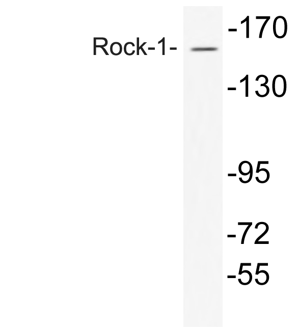
Anti-Rock-1 AntibodyA95593
ApplicationsWestern Blot, ELISA, ImmunoHistoChemistry
ReactivityHuman, Mouse, Rat
- SizePrice

Product group Antibodies
Anti-ROCK1 Antibody Picoband(r)A00722-4-CARRIER-FREE
ApplicationsFlow Cytometry, Western Blot, ELISA
ReactivityHuman, Monkey, Mouse, Rat
TargetROCK1
- SizePrice
Product group Antibodies
Anti-ROCK1 Antibody144-01008
ApplicationsImmunoPrecipitation, Western Blot, ImmunoHistoChemistry
ReactivityHuman, Mouse, Rat
TargetROCK1
- SizePrice
Product group Antibodies
References
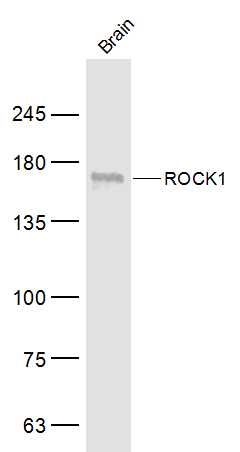
ROCK1 Polyclonal AntibodyBS-1166R
ApplicationsImmunoFluorescence, Western Blot, ELISA, ImmunoCytoChemistry, ImmunoHistoChemistry, ImmunoHistoChemistry Frozen, ImmunoHistoChemistry Paraffin
ReactivityHuman, Mouse, Rabbit, Rat
TargetROCK1
- SizePrice
Product group Antibodies
ROCK1 AntibodyCSB-PA004017
ApplicationsWestern Blot, ELISA, ImmunoHistoChemistry
ReactivityHuman, Monkey, Mouse, Rat
TargetROCK1
- SizePrice
Product group Antibodies
ROCK1 Polyclonal AntibodyCAC15747
ApplicationsImmunoFluorescence, Western Blot, ELISA, ImmunoHistoChemistry
TargetROCK1
- SizePrice
Product group Antibodies

Rho Kinase / ROCK1 AntibodyLS-C408278
ApplicationsImmunoPrecipitation, Western Blot, ImmunoHistoChemistry
ReactivityHuman, Mouse, Rat
TargetROCK1
- SizePrice

Product group Antibodies
Anti-ROCK1 AntibodyHPA007567
ApplicationsWestern Blot, ImmunoHistoChemistry
ReactivityHuman, Mouse, Rat
TargetROCK1
- SizePrice