


Immunohistochemical staining of human placenta shows strong cytoplasmic positivity in trophoblastic cells.
Immunohistochemical staining of human placenta shows strong cytoplasmic positivity in trophoblastic cells.
Anti-RPAIN Antibody
HPA023924
ApplicationsImmunoHistoChemistry
Product group Antibodies
ReactivityHuman
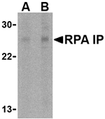
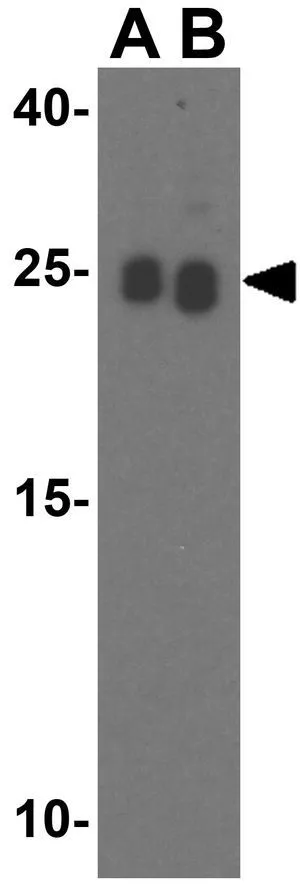
TargetRPAIN
Overview
- SupplierAtlas Antibodies
- Product NameAnti-RPAIN Antibody
- Delivery Days Customer4
- ApplicationsImmunoHistoChemistry
- CertificationResearch Use Only
- ClonalityPolyclonal
- ConjugateUnconjugated
- Gene ID84268
- Target nameRPAIN
- Target descriptionRPA interacting protein
- Target synonymsHRIP, RIP, RPA-interacting protein, RAP interaction protein, nuclear transporter
- HostRabbit
- IsotypeIgG
- Scientific DescriptionRecombinant Protein Epitope Signature Tag (PrEST) antigen sequence
- ReactivityHuman
- Storage Instruction-20°C,2°C to 8°C
- UNSPSC41116161
Datasheet
MSDS
Related products
Product group Antibodies
Anti-RPAIN (N-term) Antibody102-22389
ApplicationsWestern Blot
TargetRPAIN
- SizePrice
Product group Antibodies
RPAIN AntibodyCSB-PA020093GA01HU
ApplicationsWestern Blot, ELISA, ImmunoHistoChemistry
ReactivityHuman
TargetRPAIN
- SizePrice
Product group Antibodies
ApplicationsWestern Blot, ELISA, ImmunoHistoChemistry
ReactivityHuman, Mouse, Rat
- SizePrice
Product group Antibodies

IHC-plus(tm) RPAIN AntibodyLS-B1396
ApplicationsWestern Blot, ELISA, ImmunoHistoChemistry, ImmunoHistoChemistry Paraffin
ReactivityHuman, Mouse, Rat
TargetRPAIN
- SizePrice

Product group Antibodies
Anti-RPAIN AntibodyHPA031526
ApplicationsImmunoCytoChemistry, ImmunoHistoChemistry
ReactivityHuman
TargetRPAIN
- SizePrice
Product group Antibodies
RPA Interacting Protein antibodyGTX85485
ApplicationsWestern Blot, ELISA
ReactivityHuman, Mouse, Rat
TargetRPAIN
- SizePrice

Product group Antibodies
Anti-RPAIN Antibody Picoband(r)A11439-1-CARRIER-FREE
ApplicationsFlow Cytometry, Western Blot, ELISA
ReactivityHuman, Mouse
TargetRPAIN
- SizePrice