


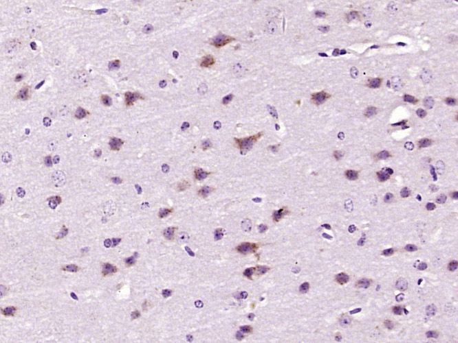
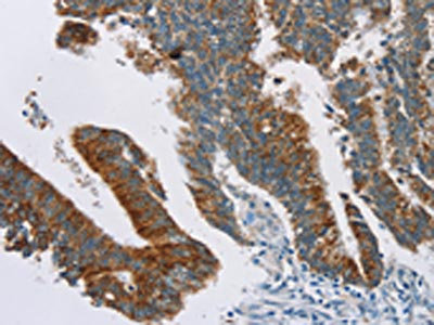
Immunohistochemical staining of human prostate shows strong cytoplasmic and membranous positivity in glandular cells.

Immunohistochemical staining of human prostate shows strong cytoplasmic and membranous positivity in glandular cells.
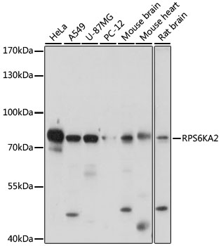
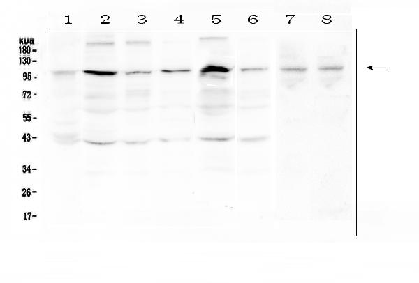
Anti-RPS6KA2 Antibody
HPA045061
ApplicationsImmunoCytoChemistry, ImmunoHistoChemistry
Product group Antibodies
ReactivityHuman
TargetRPS6KA2
Overview
- SupplierAtlas Antibodies
- Product NameAnti-RPS6KA2 Antibody
- Delivery Days Customer4
- ApplicationsImmunoCytoChemistry, ImmunoHistoChemistry
- CertificationResearch Use Only
- ClonalityPolyclonal
- ConjugateUnconjugated
- Gene ID6196
- Target nameRPS6KA2
- Target descriptionribosomal protein S6 kinase A2
- Target synonymsHU-2, MAPKAPK1C, RSK, RSK3, S6K-alpha, S6K-alpha2, p90-RSK3, p90RSK2, pp90RSK3, ribosomal protein S6 kinase alpha-2, MAP kinase-activated protein kinase 1c, MAPK-activated protein kinase 1c, mitogen-activated protein kinase-activated protein kinase 1C, ribosomal S6 kinase 3, ribosomal protein S6 kinase, 90kDa, polypeptide 2
- HostRabbit
- IsotypeIgG
- Protein IDQ15349
- Protein NameRibosomal protein S6 kinase alpha-2
- Scientific DescriptionRecombinant Protein Epitope Signature Tag (PrEST) antigen sequence
- ReactivityHuman
- Storage Instruction-20°C,2°C to 8°C
- UNSPSC41116161
Datasheet
MSDS
Related products
Product group Antibodies
Anti-RSK3 AntibodyA91326
ApplicationsImmunoFluorescence, Western Blot, ImmunoCytoChemistry, ImmunoHistoChemistry
ReactivityHuman, Mouse, Rat
- SizePrice
Product group Antibodies
Anti-RSK3/RPS6KA2 Antibody Picoband(r)A06142-1-CARRIER-FREE
ApplicationsImmunoFluorescence, Western Blot, ELISA, ImmunoCytoChemistry, ImmunoHistoChemistry
ReactivityHuman, Mouse, Rat
TargetRPS6KA2
- SizePrice
Product group Antibodies
Anti-RPS6KA2 Antibody144-64269
ApplicationsImmunoFluorescence, Western Blot, ImmunoHistoChemistry
ReactivityHuman, Mouse, Rat
TargetRPS6KA2
- SizePrice
Product group Antibodies
ApplicationsImmunoFluorescence, Western Blot, ELISA, ImmunoCytoChemistry, ImmunoHistoChemistry, ImmunoHistoChemistry Frozen, ImmunoHistoChemistry Paraffin
ReactivityBovine, Canine, Equine, Human, Mouse, Porcine, Rabbit, Rat
TargetRPS6KA2
- SizePrice
Product group Antibodies

Goat anti-RPS6KA2EB08865
ApplicationsWestern Blot, ELISA
ReactivityEquine, Human, Mouse, Rat
TargetRPS6KA2
- SizePrice
Product group Antibodies
Rps6Ka2 Polyclonal AntibodyCAC11212
ApplicationsImmunoFluorescence, ELISA, ImmunoHistoChemistry
TargetRPS6KA2
- SizePrice
Product group Antibodies
RPS6KA2 AntibodyCSB-PA035961
ApplicationsELISA, ImmunoHistoChemistry
ReactivityHuman, Mouse
TargetRPS6KA2
- SizePrice
Product group Antibodies

RPS6KA2 / RSK3 AntibodyLS-C406739
ApplicationsELISA, ImmunoHistoChemistry
ReactivityHuman, Mouse
TargetRPS6KA2
- SizePrice