

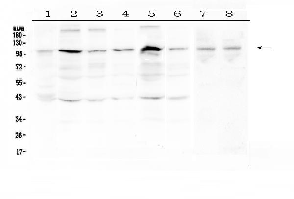
Figure 1. Western blot analysis of RSK3 using anti-RSK3 antibody (A06142-1). Electrophoresis was performed on a 5-20% SDS-PAGE gel at 70V (Stacking gel) / 90V (Resolving gel) for 2-3 hours. The sample well of each lane was loaded with 50ug of sample under reducing conditions. Lane 1: human placenta tissue lysates, Lane 2: human Hela whole cell lysate, Lane 3: human PC-3 whole cell lysate, Lane 4: human A431 whole cell lysate, Lane 5: human K562 whole cell lysate, Lane 6: human PANC-1 whole cell lysate, Lane 7: rat testis tissue lysates, Lane 8: mouse testis tissue lysates. After Electrophoresis, proteins were transferred to a Nitrocellulose membrane at 150mA for 50-90 minutes. Blocked the membrane with 5% Non-fat Milk/ TBS for 1.5 hour at RT. The membrane was incubated with rabbit anti-RSK3 antigen affinity purified polyclonal antibody (Catalog # A06142-1) at 0.5 microg/mL overnight at 4°C, then washed with TBS-0.1%Tween 3 times with 5 minutes each and probed with a goat anti-rabbit IgG-HRP secondary antibody at a dilution of 1:10000 for 1.5 hour at RT. The signal is developed using an Enhanced Chemiluminescent detection (ECL) kit (Catalog # EK1002) with Tanon 5200 system. A specific band was detected for RSK3 at approximately 90-100KD. The expected band size for RSK3 is at 83KD.
Figure 1. Western blot analysis of RSK3 using anti-RSK3 antibody (A06142-1). Electrophoresis was performed on a 5-20% SDS-PAGE gel at 70V (Stacking gel) / 90V (Resolving gel) for 2-3 hours. The sample well of each lane was loaded with 50ug of sample under reducing conditions. Lane 1: human placenta tissue lysates, Lane 2: human Hela whole cell lysate, Lane 3: human PC-3 whole cell lysate, Lane 4: human A431 whole cell lysate, Lane 5: human K562 whole cell lysate, Lane 6: human PANC-1 whole cell lysate, Lane 7: rat testis tissue lysates, Lane 8: mouse testis tissue lysates. After Electrophoresis, proteins were transferred to a Nitrocellulose membrane at 150mA for 50-90 minutes. Blocked the membrane with 5% Non-fat Milk/ TBS for 1.5 hour at RT. The membrane was incubated with rabbit anti-RSK3 antigen affinity purified polyclonal antibody (Catalog # A06142-1) at 0.5 microg/mL overnight at 4°C, then washed with TBS-0.1%Tween 3 times with 5 minutes each and probed with a goat anti-rabbit IgG-HRP secondary antibody at a dilution of 1:10000 for 1.5 hour at RT. The signal is developed using an Enhanced Chemiluminescent detection (ECL) kit (Catalog # EK1002) with Tanon 5200 system. A specific band was detected for RSK3 at approximately 90-100KD. The expected band size for RSK3 is at 83KD.
Anti-RSK3/RPS6KA2 Antibody Picoband(r)
A06142-1-CARRIER-FREE
ApplicationsImmunoFluorescence, Western Blot, ELISA, ImmunoCytoChemistry, ImmunoHistoChemistry
Product group Antibodies
ReactivityHuman, Mouse, Rat
TargetRPS6KA2
Overview
- SupplierBoster Bio
- Product NameAnti-RSK3/RPS6KA2 Antibody Picoband(r)
- Delivery Days Customer9
- ApplicationsImmunoFluorescence, Western Blot, ELISA, ImmunoCytoChemistry, ImmunoHistoChemistry
- CertificationResearch Use Only
- ClonalityPolyclonal
- Concentration500 ug/ml
- Gene ID6196
- Target nameRPS6KA2
- Target descriptionribosomal protein S6 kinase A2
- Target synonymsHU-2, MAPKAPK1C, RSK, RSK3, S6K-alpha, S6K-alpha2, p90-RSK3, p90RSK2, pp90RSK3, ribosomal protein S6 kinase alpha-2, MAP kinase-activated protein kinase 1c, MAPK-activated protein kinase 1c, mitogen-activated protein kinase-activated protein kinase 1C, ribosomal S6 kinase 3, ribosomal protein S6 kinase, 90kDa, polypeptide 2
- HostRabbit
- IsotypeIgG
- Protein IDQ15349
- Protein NameRibosomal protein S6 kinase alpha-2
- Scientific DescriptionBoster Bio Anti-RSK3/RPS6KA2 Antibody Picoband® catalog # A06142-1. Tested in ELISA, IF, IHC, ICC, WB applications. This antibody reacts with Human, Mouse, Rat. The brand Picoband indicates this is a premium antibody that guarantees superior quality, high affinity, and strong signals with minimal background in Western blot applications. Only our best-performing antibodies are designated as Picoband, ensuring unmatched performance.
- ReactivityHuman, Mouse, Rat
- Storage Instruction-20°C,2°C to 8°C
- UNSPSC12352203
Related products
Product group Antibodies
Anti-RSK3 AntibodyA91326
ApplicationsImmunoFluorescence, Western Blot, ImmunoCytoChemistry, ImmunoHistoChemistry
ReactivityHuman, Mouse, Rat
- SizePrice
Product group Antibodies
Anti-RPS6KA2 Antibody144-64269
ApplicationsImmunoFluorescence, Western Blot, ImmunoHistoChemistry
ReactivityHuman, Mouse, Rat
TargetRPS6KA2
- SizePrice
Product group Antibodies
ApplicationsImmunoFluorescence, Western Blot, ELISA, ImmunoCytoChemistry, ImmunoHistoChemistry, ImmunoHistoChemistry Frozen, ImmunoHistoChemistry Paraffin
ReactivityBovine, Canine, Equine, Human, Mouse, Porcine, Rabbit, Rat
TargetRPS6KA2
- SizePrice
Product group Antibodies

Goat anti-RPS6KA2EB08865
ApplicationsWestern Blot, ELISA
ReactivityEquine, Human, Mouse, Rat
TargetRPS6KA2
- SizePrice
Product group Antibodies
Rps6Ka2 Polyclonal AntibodyCAC11212
ApplicationsImmunoFluorescence, ELISA, ImmunoHistoChemistry
TargetRPS6KA2
- SizePrice
Product group Antibodies
RPS6KA2 AntibodyCSB-PA035961
ApplicationsELISA, ImmunoHistoChemistry
ReactivityHuman, Mouse
TargetRPS6KA2
- SizePrice
Product group Antibodies

RPS6KA2 / RSK3 AntibodyLS-C406739
ApplicationsELISA, ImmunoHistoChemistry
ReactivityHuman, Mouse
TargetRPS6KA2
- SizePrice

Product group Antibodies
Anti-RPS6KA2 AntibodyHPA045061
ApplicationsImmunoCytoChemistry, ImmunoHistoChemistry
ReactivityHuman
TargetRPS6KA2
- SizePrice
![Various whole cell extracts (30 μg) were separated by 7.5% SDS-PAGE, and the membrane was blotted with RSK3 antibody [N2C1], Internal (GTX111071) diluted at 1:1000. The HRP-conjugated anti-rabbit IgG antibody (GTX213110-01) was used to detect the primary antibody.](https://www.genetex.com/upload/website/prouct_img/normal/GTX111071/GTX111071_40044_20190118_WB_M_w_23060500_206.webp)
Product group Antibodies
RSK3 antibody [N2C1], InternalGTX111071
ApplicationsWestern Blot
ReactivityHuman, Mouse
TargetRPS6KA2
- SizePrice