

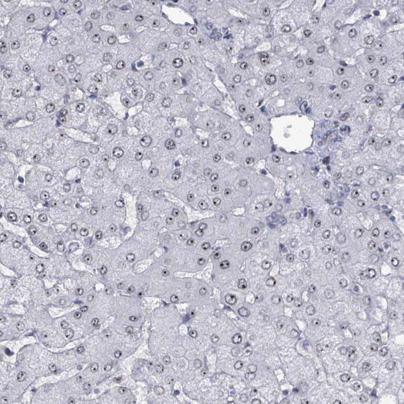
Immunohistochemical staining of human liver shows strong nucleolar positivity in hepatocytes.

Immunohistochemical staining of human liver shows strong nucleolar positivity in hepatocytes.
Anti-RRN3 Antibody
HPA049837
ApplicationsImmunoCytoChemistry
Product group Antibodies
ReactivityHuman
TargetRRN3
Overview
- SupplierAtlas Antibodies
- Product NameAnti-RRN3 Antibody
- Delivery Days Customer4
- ApplicationsImmunoCytoChemistry
- CertificationResearch Use Only
- ClonalityPolyclonal
- ConjugateUnconjugated
- Gene ID54700
- Target nameRRN3
- Target descriptionRNA polymerase I transcription factor RRN3
- Target synonymsA-270G1.2, TIFIA, RNA polymerase I-specific transcription initiation factor RRN3, RRN3 RNA polymerase I transcription factor homolog, RRN3 homolog, RNA polymerase I transcription factor, TIF-IA, transcription initiation factor IA, transcription initiation factor TIF-IA
- HostRabbit
- IsotypeIgG
- Protein IDQ9NYV6
- Protein NameRNA polymerase I-specific transcription initiation factor RRN3
- Scientific DescriptionRecombinant Protein Epitope Signature Tag (PrEST) antigen sequence
- ReactivityHuman
- Storage Instruction-20°C,2°C to 8°C
- UNSPSC41116161
Datasheet
MSDS
Related products
Product group Antibodies
Anti-RRN3 (Center) Antibody102-22687
ApplicationsWestern Blot
TargetRRN3
- SizePrice
Product group Antibodies
ApplicationsWestern Blot, ImmunoHistoChemistry
ReactivityHuman
- SizePrice
Product group Antibodies

TIF-IA / RRN3 AntibodyLS-C813526
ApplicationsWestern Blot, ELISA
ReactivityHuman, Rat
TargetRRN3
- SizePrice
Product group Antibodies
Phospho-RRN3 (S649) AntibodyCSB-PA030142
ApplicationsWestern Blot, ELISA, ImmunoHistoChemistry
ReactivityHuman
TargetRRN3
- SizePrice
Product group Antibodies
RRN3 antibody [N1N3]GTX117115
ApplicationsWestern Blot
ReactivityHuman
TargetRRN3
- SizePrice

Product group Antibodies
Anti-RRN3 Antibody Picoband(r)A05439-2-CARRIER-FREE
ApplicationsFlow Cytometry, Western Blot, ELISA
ReactivityHuman
TargetRRN3
- SizePrice