


Immunofluorescent staining of human cell line U-2 OS shows localization to nucleus, nucleoli & cytosol.

Immunofluorescent staining of human cell line U-2 OS shows localization to nucleus, nucleoli & cytosol.
Anti-RRP8 Antibody
HPA057562
ApplicationsWestern Blot, ImmunoCytoChemistry
Product group Antibodies
ReactivityHuman
TargetRRP8
Overview
- SupplierAtlas Antibodies
- Product NameAnti-RRP8 Antibody
- Delivery Days Customer4
- ApplicationsWestern Blot, ImmunoCytoChemistry
- CertificationResearch Use Only
- ClonalityPolyclonal
- ConjugateUnconjugated
- Gene ID23378
- Target nameRRP8
- Target descriptionribosomal RNA processing 8
- Target synonymsKIAA0409, NML, ribosomal RNA-processing protein 8, RRP8 methyltransferase homolog, cerebral protein 1, nucleomethylin, ribosomal RNA processing 8, methyltransferase, homolog
- HostRabbit
- IsotypeIgG
- Protein IDO43159
- Protein NameRibosomal RNA-processing protein 8
- Scientific DescriptionRecombinant Protein Epitope Signature Tag (PrEST) antigen sequence
- ReactivityHuman
- Storage Instruction-20°C,2°C to 8°C
- UNSPSC41116161
Datasheet
MSDS
Related products
Product group Antibodies
RRP8 AntibodyCSB-PA008360
ApplicationsWestern Blot, ELISA, ImmunoHistoChemistry
ReactivityHuman
TargetRRP8
- SizePrice

Product group Antibodies
Anti-RRP8 Antibody Picoband(r)A09950-2-CARRIER-FREE
ApplicationsFlow Cytometry, ImmunoFluorescence, Western Blot, ELISA, ImmunoCytoChemistry
ReactivityHuman, Mouse, Rat
TargetRRP8
- SizePrice

Product group Antibodies
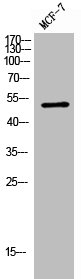
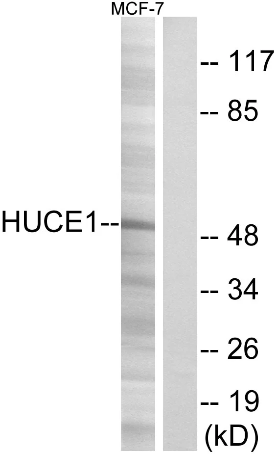
Anti-HUCE1 AntibodyA101040
ApplicationsWestern Blot, ELISA
ReactivityHuman
- SizePrice
Product group Antibodies

RRP8 AntibodyLS-C830159
ApplicationsELISA, ImmunoHistoChemistry
ReactivityHuman, Mouse, Rat
TargetRRP8
- SizePrice

Product group Antibodies
Anti-RRP8 AntibodyHPA038487
ApplicationsImmunoCytoChemistry, ImmunoHistoChemistry
ReactivityHuman
TargetRRP8
- SizePrice
Product group Antibodies
Anti-RRP8 AntibodyHPA038487
ApplicationsImmunoCytoChemistry, ImmunoHistoChemistry
ReactivityHuman
TargetRRP8
- SizePrice
Product group Antibodies
HUCE1 antibodyGTX87185
ApplicationsWestern Blot
ReactivityHuman
TargetRRP8
- SizePrice