

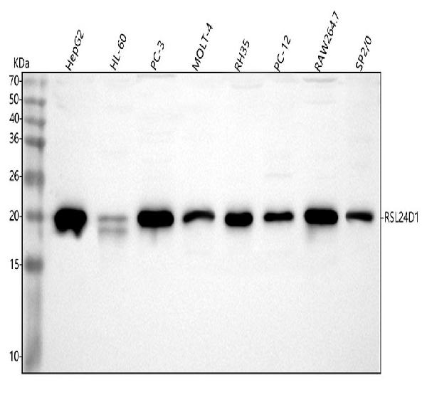
Figure 1. Western blot analysis of RSL24D1 using anti-RSL24D1 antibody (A14856-1). Electrophoresis was performed on a 5-20% SDS-PAGE gel at 70V (Stacking gel) / 90V (Resolving gel) for 2-3 hours. The sample well of each lane was loaded with 30 ug of sample under reducing conditions. Lane 1: human HepG2 whole cell lysates, Lane 2: human HL-60 whole cell lysates, Lane 3: human PC-3 whole cell lysates, Lane 4: human MOLT-4 whole cell lysates, Lane 5: rat RH35 whole cell lysates, Lane 6: rat PC-12 whole cell lysates, Lane 7: mouse RAW264.7 whole cell lysates, Lane 8: mouse SP2/0 whole cell lysates. After electrophoresis, proteins were transferred to a nitrocellulose membrane at 150 mA for 50-90 minutes. Blocked the membrane with 5% non-fat milk/TBS for 1.5 hour at RT. The membrane was incubated with rabbit anti-RSL24D1 antigen affinity purified polyclonal antibody (Catalog # A14856-1) at 0.25 microg/mL overnight at 4°C, then washed with TBS-0.1%Tween 3 times with 5 minutes each and probed with a goat anti-rabbit IgG-HRP secondary antibody at a dilution of 1:5000 for 1.5 hour at RT. The signal is developed using an Enhanced Chemiluminescent detection (ECL) kit (Catalog # EK1002) with Tanon 5200 system. A specific band was detected for RSL24D1 at approximately 20 kDa. The expected band size for RSL24D1 is at 20 kDa.
. RSL24D1 was detected in an immunocytochemical section of A549 cells. Enzyme antigen retrieval was performed using IHC enzyme antigen retrieval reagent (AR0022) for 15 mins. The cells were blocked with 10% goat serum. And then incubated with 5 microg/mL rabbit anti-RSL24D1 Antibody (A14856-1) overnight at 4°C. Cy3 Conjugated Goat Anti-Rabbit IgG (BA1032) was used as secondary antibody at 1:500 dilution and incubated for 30 minutes at 37°C. The section was counterstained with DAPI. Visualize using a fluorescence microscope and filter sets appropriate for the label used.")
. Overlay histogram showing JK cells stained with A14856-1 (Blue line). To facilitate intracellular staining, cells were fixed with 4% paraformaldehyde and permeabilized with permeabilization buffer. The cells were blocked with 10% normal goat serum. And then incubated with rabbit anti-RSL24D1 Antibody (A14856-1, 1 microg/1x106 cells) for 30 min at 20°C. DyLight®488 conjugated goat anti-rabbit IgG (BA1127, 5-10 microg/1x106 cells) was used as secondary antibody for 30 minutes at 20°C. Isotype control antibody (Green line) was rabbit IgG (1 microg/1x106) used under the same conditions. Unlabelled sample (Red line) was also used as a control.")
Figure 1. Western blot analysis of RSL24D1 using anti-RSL24D1 antibody (A14856-1). Electrophoresis was performed on a 5-20% SDS-PAGE gel at 70V (Stacking gel) / 90V (Resolving gel) for 2-3 hours. The sample well of each lane was loaded with 30 ug of sample under reducing conditions. Lane 1: human HepG2 whole cell lysates, Lane 2: human HL-60 whole cell lysates, Lane 3: human PC-3 whole cell lysates, Lane 4: human MOLT-4 whole cell lysates, Lane 5: rat RH35 whole cell lysates, Lane 6: rat PC-12 whole cell lysates, Lane 7: mouse RAW264.7 whole cell lysates, Lane 8: mouse SP2/0 whole cell lysates. After electrophoresis, proteins were transferred to a nitrocellulose membrane at 150 mA for 50-90 minutes. Blocked the membrane with 5% non-fat milk/TBS for 1.5 hour at RT. The membrane was incubated with rabbit anti-RSL24D1 antigen affinity purified polyclonal antibody (Catalog # A14856-1) at 0.25 microg/mL overnight at 4°C, then washed with TBS-0.1%Tween 3 times with 5 minutes each and probed with a goat anti-rabbit IgG-HRP secondary antibody at a dilution of 1:5000 for 1.5 hour at RT. The signal is developed using an Enhanced Chemiluminescent detection (ECL) kit (Catalog # EK1002) with Tanon 5200 system. A specific band was detected for RSL24D1 at approximately 20 kDa. The expected band size for RSL24D1 is at 20 kDa.
Anti-RSL24D1 Antibody Picoband(r)
A14856-1-CARRIER-FREE
ApplicationsFlow Cytometry, ImmunoFluorescence, Western Blot, ELISA, ImmunoCytoChemistry
Product group Antibodies
ReactivityHuman, Mouse, Rat
TargetRSL24D1
Overview
- SupplierBoster Bio
- Product NameAnti-RSL24D1 Antibody Picoband(r)
- Delivery Days Customer9
- ApplicationsFlow Cytometry, ImmunoFluorescence, Western Blot, ELISA, ImmunoCytoChemistry
- CertificationResearch Use Only
- ClonalityPolyclonal
- Concentration500 ug/ml
- Gene ID51187
- Target nameRSL24D1
- Target descriptionribosomal L24 domain containing 1
- Target synonymsC15orf15, HRP-L30-iso, L30, RLP24, RPL24, RPL24L, TVAS3, probable ribosome biogenesis protein RLP24, 60S ribosomal protein L30 isolog, homolog of yeast ribosomal like protein 24, my024 protein, ribosomal L24 domain-containing protein 1
- HostRabbit
- IsotypeIgG
- Protein IDQ9UHA3
- Protein NameProbable ribosome biogenesis protein RLP24
- Scientific DescriptionBoster Bio Anti-RSL24D1 Antibody Picoband® catalog # A14856-1. Tested in WB, ICC/IF, Flow Cytometry, ELISA applications. This antibody reacts with Human, Mouse, Rat. The brand Picoband indicates this is a premium antibody that guarantees superior quality, high affinity, and strong signals with minimal background in Western blot applications. Only our best-performing antibodies are designated as Picoband, ensuring unmatched performance.
- ReactivityHuman, Mouse, Rat
- Storage Instruction-20°C,2°C to 8°C
- UNSPSC12352203
Related products
Product group Antibodies
Anti-RSL24D1 AntibodyA308676
ApplicationsWestern Blot
ReactivityHuman, Mouse
- SizePrice
Product group Antibodies
Anti-RSL24D1 Antibody144-65091
ApplicationsWestern Blot
ReactivityHuman, Mouse
TargetRSL24D1
- SizePrice

Product group Antibodies
Anti-RSL24D1 AntibodyHPA062724
ApplicationsWestern Blot, ImmunoCytoChemistry, ImmunoHistoChemistry
ReactivityHuman
TargetRSL24D1
- SizePrice
Product group Antibodies
C15ORF15 antibody, InternalGTX45009
ApplicationsWestern Blot
ReactivityHuman
TargetRSL24D1
- SizePrice
Product group Antibodies
Anti-RSL24D1 AntibodyCAB17700
ApplicationsWestern Blot, ELISA
ReactivityHuman
TargetRSL24D1
- SizePrice
Product group Antibodies

ApplicationsImmunoFluorescence, Western Blot, ELISA, ImmunoHistoChemistry, ImmunoHistoChemistry Paraffin
ReactivityHuman
TargetRSL24D1
- SizePrice